Gallery: Early immunofluorescence
How did immunofluorescence begin? Who was the first scientist to use fluorescently labeled antibodies to stain cells under a microscope?
The answer to this is pretty clear. Albert Coons of Harvard Medical School, in two papers from 1942 and 1950. In this 1962 speech (published in JAMA) Wesley Spink of the University of Minnesota describes Coons’s accomplishments.
The first time fluorescent molecules were attached to antibodies was in the early 1930s, as described in a brief letter to Nature by John Marrack (1934). But at this point the antibodies are being used in suspension, for serological experiments similar to those that detect bacteria by agglutination. In this case the bacteria can be measured colorimetrically by mixing them with fluorescently labeled antibody, washing off nonspecific antibodies, and seeing if the bacteria turn pink.
In fact, here’s the entire article.
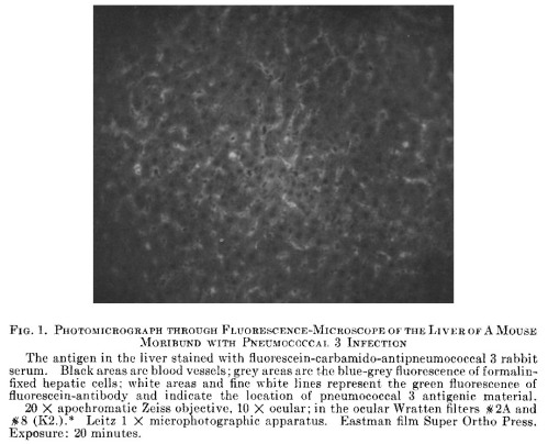
In The Demonstration of Pneumococcal Antigen in Tissues by Means of Fluorescent Antibody (1942), Coons and associates showed that fluorescein isocyanate (FIC) could be attached to antibodies and used to stain thin tissue sections on slides, in the same way many histological stains had been used in the past. Rabbits were immunized with pneumococcus, and their serum was conjugated with FIC. This was used to visualize the bacteria in the liver of a mouse with severe pneumococcus infection. The first immunofluorescence image ever published, I believe, was this 20-minute (!) exposure.
* * *
But the fluorescent antibody technique didn’t take off until Coons and Kaplan published the sequel eight years later. I can’t find any papers from the 1940s that cite the Coons paper and actually include fluorescent images of their own.
The pivotal 1950 paper is entitled Localization of Antigen in Tissue Cells: II: Improvements in a Method for the Detection of Antigen by Means of Fluorescent Antibody. Kind of like Rambo III, the numbering of this title is odd because there was no Localization of Antigen in Tissue Cells Part I. Instead, it’s explained that “the first paper in this series was entitled ‘The Demonstration of Pneumococcal Antigen in Tissues by Means of Fluorescent Antibody'”, meaning that this is a sequel 8 years in the making.
And what a sequel! Over 2,100 citations, according to Google Scholar’s notoriously inflated algorithm. Meanwhile PubMed’s claim of 301 is a clear underestimate. It was cited a lot.
Suddenly everyone was able to use fluorescent antibodies to find out where their antigen of choice was located inside the cell, or inside the tissue, or inside the animal. Especially after 1958 when Riggs and colleagues at the University of Kansas showed how to make FITC (fluorescein isothiocyanate) conjugates, which are more stable than FIC and don’t require phosgene for their chemical synthesis. Using thiophosgene is no picnic either, but apparently it’s less deadly.
* * *
Just as early issues of the Journal of Virology are filled with papers that show “The Ultrastructure of [Name of virus]” by taking random pictures of it with an electron microscope, it seems like it was easy to get a microscopy paper published in the early 1960s. You injected rabbits with a substance, labeled their serum with fluorescein, stained some slides, and put together a manuscript called “Demonstration of [Name of substance] in [Name of tissue] by the Fluorescent Antibody Technique”.
I decided to look through some papers that cite Coons and Kaplan (1950), to find examples of early immunofluorescence. Here are a dozen images, all from at least fifty years ago.
* * *
(2) Chicken muscle fiber, stained with rabbit globulin specific for myosin (0.5 microns, 1000x). (3) A “dark medium phase contrast” image of the same slide for comparison. (Finck et al., J Biophys Biochem Cytol 1956)
—
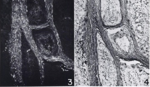
(3) Cottontail rabbit papilloma, stained with rabbit serum specific for Shope papilloma virus and goat anti-rabbit (75x). (4) An H&E stain of the same slide, to show that the virus antigens are in the keratinized region. (Mellors, Cancer Res 1960)
—
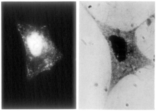
Embryonic chicken fibroblast infected for 10 hours with the Rostock strain of fowl plague virus (now known to be influenza A virus H7N1), stained with rabbit serum specific for “g antigen” (now called NP) (950x). The same cell stained by Giemsa-Wright stain, to show the nucleus. (Breitenfeld and Schäfer, Virology 1957)
—
Rat eye (lens and iris), stained with rabbit globulin specific for rat glomerulus (190x). Another section of the same eye stained with non-specific rabbit globulin. This is an example of the papers that used specific antiserum to find common antigens in seemingly unrelated tissues, in this case kidney and eye. (Roberts, Br J Ophthalmol 1957)
—
Human tissues, stained with human serum specific for blood group A or B. Yes, human serum. A volunteer of blood group A was used to get the serum against blood group B, and vice versa. (Szulman, J Exp Med 1960)
—
Group B streptococci, stained with rabbit globulin specific for group B streptococci (magnification unspecified). This paper’s total lack of negative controls is charmingly naive. (Moody et al., J Bacteriol 1958)
—
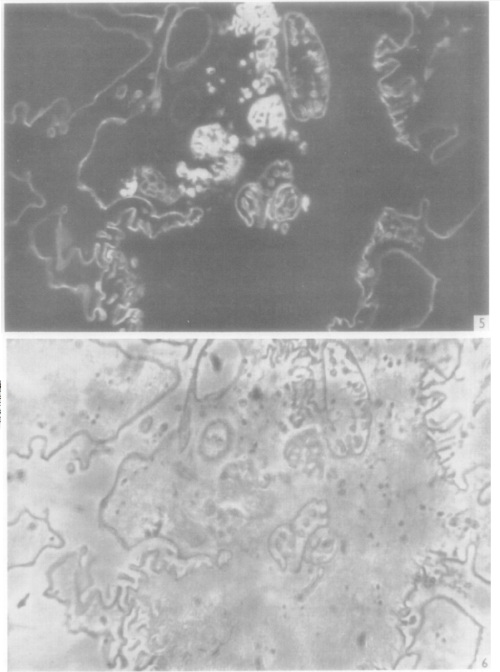
(5) Amoebae frozen during pinocytosis, with free fluorescent antibody (non-specific rabbit globulin) visible in pinocytosis vacuoles (2000x). (6) Phase-contrast view of another section of the same amoeba. (Brandt, Exp Cell Res 1958)
—
Mononuclear cells from nasal smears of ferrets infected with influenza (PR8 strain), stained with rabbit globulin specific for influenza virus (560x). Cells display a range of nuclear and cytoplasmic staining patterns. (Liu, J Exp Med 1955)
—
Cladosporium bantianum mold stained with rabbit serum specific for C. bantianum (1000x). By comparison, C. carrionii stained with serum specific for C. bantianum, to show lack of cross-reactivity. (Al-Doory and Gordon, J Bacteriol 1963)
—
Mixture of two yeast species photographed under both ultraviolet and visible light simultaneously (magnification unspecified). Saccharomyces cerevisiae (near center) glows green when stained by rabbit serum specific for S. cerevisiae. Red light serves as counterstain for other species (Pichia membranefaciens). (Kunz and Klaushofer, Appl Microbiol 1961)
—
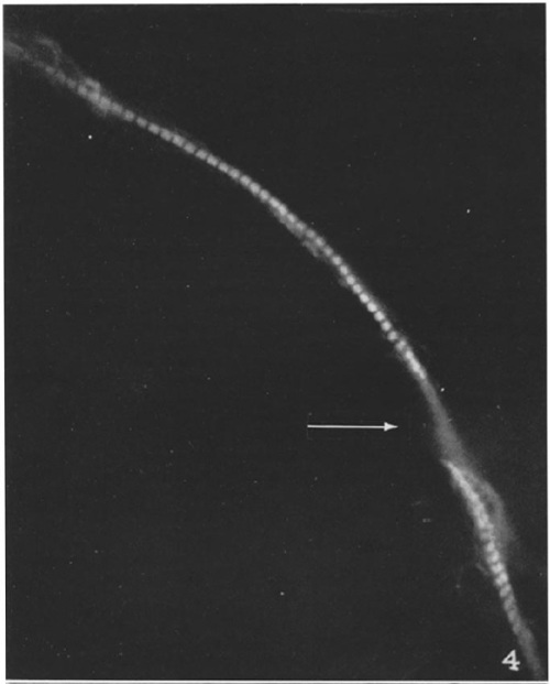
A single myoblast isolated from a stage 23 chick embryo, stained with rabbit globulin specific for myosin (1100x). The arrow indicates where the nucleus obscures the myosin. (Holtzer et al., J Biophys Biochem Cytol 1957)
—
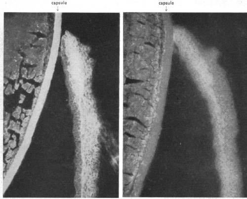
Kidney of rat injected with nephrotoxic rabbit antibodies to cause nephritis; the rabbit antibodies concentrate in the glomeruli, as seen by staining with goat globulin specific for rabbit globulin (37x and 205x). (Ortega and Mellors, J Exp Med 1956)


