Amboceptor

Microbiological, virological, bacteriological, immunological, medical, epidemiological, historical, anecdotal

Tag: antibodies

Pubmedwhack: Immuno-DTO

Today’s Pubmedwhack comes from the world of unstable metals and electron microscopy. For the definition of “Pubmedwhack”, see this earlier post.

* * *

Following the invention of immunofluorescence, scientists developed other methods for using labeled antibodies to identify certain proteins or substances under a microscope.

There’s immunohistochemistry (IHC), in which the antibodies are labeled not with a fluorescent marker, but with an enzyme which produces a visible reaction in the presence of a substrate. This is less precise, but lets you see your protein of choice under visible light while also looking at the structure of the tissue. The slides labeled this way last longer instead of being quenched by the microscope’s light source.

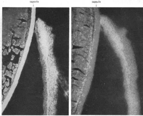
You can also use labeled antibodies to see things under the electron microscope (immuno-electron microscopy or immuno-EM). In 1960 Rifkin et al. (1) published an image of virus particles on a cell surface, labeled with ferritin-conjugated antibodies. As you can see, instead of the labeled antibodies changing the color of a region of the cell, each individual labeled antibody is visible as a “granule”.

rifkin-1960-ferritin

from Rifkin et al (1960), Nature 187:1094

This was made possible by an innovation published one year earlier, entitled Preparation of an electron-dense antibody conjugate (2). Ferritin is a small protein which just about all organisms use as an iron carrier. When “iron-loaded”, almost a quarter of its mass is iron atoms. Therefore this is an especially electron-dense molecule, visible as a dark spot under the electron microscope, as seen above. Soon, further advances let scientists see ferritin-labeled structures inside cells.

* * *

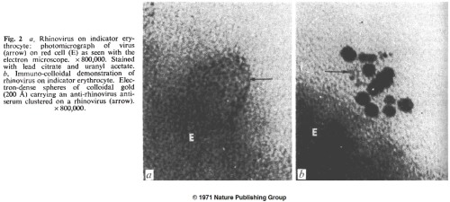
For about a decade ferritin was the label of choice for immuno-electron microscopy. Then in 1971, a new technique came along (3, 4), in which antibodies were mixed with a solution of colloidal gold until they absorbed to the metal’s surface. Gold-labeled antibodies could be separated from free antibodies by centrifugation. Immunogold is still the dominant immuno-EM staining method 40 years later.

from Faulk (1971), Nature New Biology 231:101

from Faulk et al (1971), Nature New Biology 231:101

* * *

In the 1960s, other techniques were created for immuno-EM. I have almost no EM experience and don’t know the pros and cons, but clearly there was a desire to get rid of the protein element of the electron-dense antibody label, and just attach the antibody to a metal ion. The protein was unnecessarily big, and subject to denaturation. So the 1960s also saw a lot of papers using antibodies labeled with mercury (technically the diazonium salt of tetraacetoxymercuriarsanilic acid (5) and p-(aminophenyl)-mercuric acetate (6), phrases which mean little to me).

from Zhdanov et al (1965), J Histochem Cytochem

from Zhdanov et al (1965), J Histochem Cytochem 13:684

There were also studies using antibodies labeled with heavier metals. In his long career, Ludwig Sternberger and his lab (at Johns Hopkins and elsewhere) invented several microscopy techniques, with the most important probably being the horseradish peroxidase (see original paper (7), and appreciation of it as a “citation classic”).

He also spent much of the 1960s devising improved metal-based antibody labels for electron microscopy, including immunouranium (8), immunouranium with added osmium for enhanced contrast (9), and finally immuno-diazothioether-osmium tetroxide (10), or immuno-DTO. The uranium methods seem somewhat useful, but as far as I can tell were only used by Sternberger’s own lab. Immuno-DTO in particular seemed almost unusable; they used it more than once, but a Pubmed search for “Immuno-DTO” only returns one result (11). As Sternberger himself says in a review (12) of his and other techniques:

Unfortunately, the diazotized diazothioethers are not very stable even at Dry Ice temperatures and the solid deteriorates in a few days. Therefore, it was not surprising to observe that the osmnium tetroxide-binding iower of diazothioether antibodies was unstable, even on storage in liquid nitrogen.

Oh well, it was a nice idea.

* * *

1. Rifkind RA, Hsu KC, Morgan C, Seegal BC, Knox AW, Rose HM (1960). Use of Ferritin-Conjugated Antibody to Localize Antigen by Electron Microscopy. Nature 187:1094-1095.

2. Singer SJ (1959). Preparation of an Electron-dense Antibody Conjugate. Nature 183:1523-1524.

3.Faulk WP, Taylor GM (1971). An Immunocolloid Method for the Electron Microscopy. Immunochemistry 8(11):1081-1083.

4. Faulk WP, Vyas GN, Phillips CA, Fudenberg HH, Chism K (1971). Passive Haemagglutination Test for Anti-rhinovirus Antibodies. Nature New Biology 231:101-104.

5. Pepe FA (1961). The Use of Specific Antibody in Electron Microscopy: I: Preparation of Mercury-Labeled Antibody. J Biophys Biochem Cytol 11(3):515-520.

6. Zhdanov VM, Azadova NB, Kulberg AY (1965). The Use of Antibody Labeled with an Organic Mercury Compound in Electron Microscopy. J Histochem Cytochem 13(8):694-687.

7. Sternberger LA, Hardy PH Jr, Cuculis JJ, Meyer HG (1970). The Unlabeled Antibody Enzyme Method of Immunohistochemistry: Preparation and Properties of Soluble Antigen-Antibody Complex (Horseradish Peroxidase-Antihorseradish Peroxidase) and its Use in Identification of Spirochetes. J Histochem Cytochem 18(5):315-333.

8. Donati EJ, Figge FHJ, Sternberger LA (1965). Staining of Vaccinia Antigen by Immunouranium Technique. Exp Mol Pathol 4(1):126-129.

9. Sternberger LA, Hanker JS, Donati EJ, Petrali JP, Seligman AM (1966). Method for Enhancement of Electron Microscopic Visualization of Embedded Antigen by Bridging Osmium to Uranium Antibody with Thiocarbohydrazide. J Histochem Cytochem 14(10):711-718.

10. Donati EJ, Petrali JP, Sternberger LA (1966). Formation of Vaccinia Antigen Studied by Immunouranium and Immuno-diaxothioether-osmium tetroxide Techniques. Exp Mol Pathol Apr:Suppl 3:59-74.

11. Sternberger LA, Donati EJ, Hanker JS, Seligman AM (1966). Immuno-diazothioether-osmium tetroxide (immuno-DTO) technique for staining embedded antigen in electron microscopy. Exp Mol Pathol Apr:Suppl 3:36-43.

12. Sternberger LA (1967). Electron Microscopic Immunocytochemistry: A Review. J Histochem Cytochem 15(3):139-159.

Gallery: Early immunofluorescence

How did immunofluorescence begin? Who was the first scientist to use fluorescently labeled antibodies to stain cells under a microscope?

The answer to this is pretty clear. Albert Coons of Harvard Medical School, in two papers from 1942 and 1950. In this 1962 speech (published in JAMA) Wesley Spink of the University of Minnesota describes Coons’s accomplishments.

The first time fluorescent molecules were attached to antibodies was in the early 1930s, as described in a brief letter to Nature by John Marrack (1934). But at this point the antibodies are being used in suspension, for serological experiments similar to those that detect bacteria by agglutination. In this case the bacteria can be measured colorimetrically by mixing them with fluorescently labeled antibody, washing off nonspecific antibodies, and seeing if the bacteria turn pink.

In fact, here’s the entire article.

marrack-1934-nature

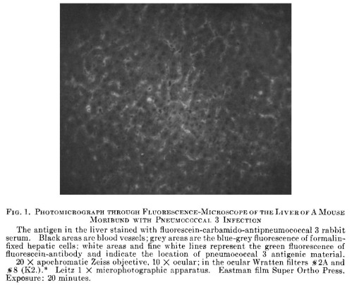
In The Demonstration of Pneumococcal Antigen in Tissues by Means of Fluorescent Antibody (1942), Coons and associates showed that fluorescein isocyanate (FIC) could be attached to antibodies and used to stain thin tissue sections on slides, in the same way many histological stains had been used in the past. Rabbits were immunized with pneumococcus, and their serum was conjugated with FIC. This was used to visualize the bacteria in the liver of a mouse with severe pneumococcus infection. The first immunofluorescence image ever published, I believe, was this 20-minute (!) exposure.

coons-1942-fluorescence

* * *

But the fluorescent antibody technique didn’t take off until Coons and Kaplan published the sequel eight years later. I can’t find any papers from the 1940s that cite the Coons paper and actually include fluorescent images of their own.

The pivotal 1950 paper is entitled Localization of Antigen in Tissue Cells: II: Improvements in a Method for the Detection of Antigen by Means of Fluorescent Antibody. Kind of like Rambo III, the numbering of this title is odd because there was no Localization of Antigen in Tissue Cells Part I. Instead, it’s explained that “the first paper in this series was entitled ‘The Demonstration of Pneumococcal Antigen in Tissues by Means of Fluorescent Antibody'”, meaning that this is a sequel 8 years in the making.

And what a sequel! Over 2,100 citations, according to Google Scholar’s notoriously inflated algorithm. Meanwhile PubMed’s claim of 301 is a clear underestimate. It was cited a lot.

Suddenly everyone was able to use fluorescent antibodies to find out where their antigen of choice was located inside the cell, or inside the tissue, or inside the animal. Especially after 1958 when Riggs and colleagues at the University of Kansas showed how to make FITC (fluorescein isothiocyanate) conjugates, which are more stable than FIC and don’t require phosgene for their chemical synthesis. Using thiophosgene is no picnic either, but apparently it’s less deadly.

* * *

Just as early issues of the Journal of Virology are filled with papers that show “The Ultrastructure of [Name of virus]” by taking random pictures of it with an electron microscope, it seems like it was easy to get a microscopy paper published in the early 1960s. You injected rabbits with a substance, labeled their serum with fluorescein, stained some slides, and put together a manuscript called “Demonstration of [Name of substance] in [Name of tissue] by the Fluorescent Antibody Technique”.

I decided to look through some papers that cite Coons and Kaplan (1950), to find examples of early immunofluorescence. Here are a dozen images, all from at least fifty years ago.

* * *

finck-chicken-myosin-1956

(2) Chicken muscle fiber, stained with rabbit globulin specific for myosin (0.5 microns, 1000x). (3) A “dark medium phase contrast” image of the same slide for comparison. (Finck et al., J Biophys Biochem Cytol 1956)

shope-papilloma-virus-mellors-1960

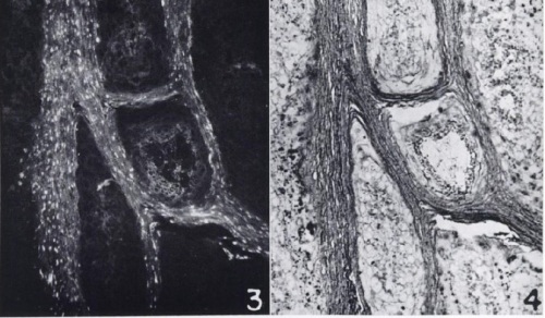
(3) Cottontail rabbit papilloma, stained with rabbit serum specific for Shope papilloma virus and goat anti-rabbit (75x). (4) An H&E stain of the same slide, to show that the virus antigens are in the keratinized region. (Mellors, Cancer Res 1960)

avian-flu-breitenfeld-1957

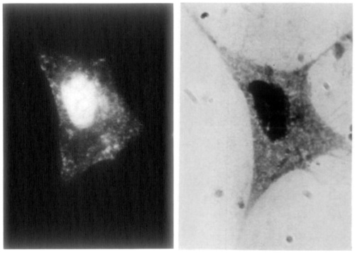
Embryonic chicken fibroblast infected for 10 hours with the Rostock strain of fowl plague virus (now known to be influenza A virus H7N1), stained with rabbit serum specific for “g antigen” (now called NP) (950x). The same cell stained by Giemsa-Wright stain, to show the nucleus. (Breitenfeld and Schäfer, Virology 1957)

roberts-1957-rat-eye

Rat eye (lens and iris), stained with rabbit globulin specific for rat glomerulus (190x). Another section of the same eye stained with non-specific rabbit globulin. This is an example of the papers that used specific antiserum to find common antigens in seemingly unrelated tissues, in this case kidney and eye. (Roberts, Br J Ophthalmol 1957)

blood-groups-a-b-szulman-1960

Human tissues, stained with human serum specific for blood group A or B. Yes, human serum. A volunteer of blood group A was used to get the serum against blood group B, and vice versa. (Szulman, J Exp Med 1960)

GBS-Moody-1960

Group B streptococci, stained with rabbit globulin specific for group B streptococci (magnification unspecified). This paper’s total lack of negative controls is charmingly naive. (Moody et al., J Bacteriol 1958)

amoeba-brandt-1958

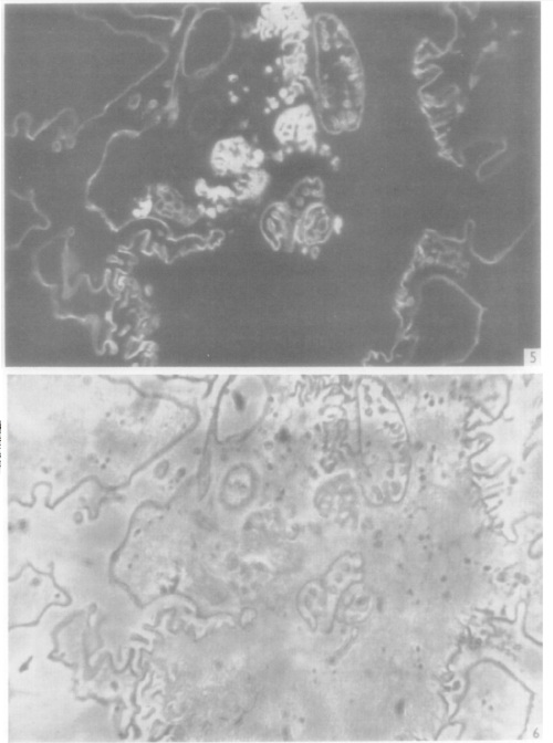
(5) Amoebae frozen during pinocytosis, with free fluorescent antibody (non-specific rabbit globulin) visible in pinocytosis vacuoles (2000x). (6) Phase-contrast view of another section of the same amoeba. (Brandt, Exp Cell Res 1958)

ferret-influenza-liu-1955

Mononuclear cells from nasal smears of ferrets infected with influenza (PR8 strain), stained with rabbit globulin specific for influenza virus (560x). Cells display a range of nuclear and cytoplasmic staining patterns. (Liu, J Exp Med 1955)

cladosporium-aldoory-1963

Cladosporium bantianum mold stained with rabbit serum specific for C. bantianum (1000x). By comparison, C. carrionii stained with serum specific for C. bantianum, to show lack of cross-reactivity. (Al-Doory and Gordon, J Bacteriol 1963)

yeast-kunz-1961

Mixture of two yeast species photographed under both ultraviolet and visible light simultaneously (magnification unspecified). Saccharomyces cerevisiae (near center) glows green when stained by rabbit serum specific for S. cerevisiae. Red light serves as counterstain for other species (Pichia membranefaciens). (Kunz and Klaushofer, Appl Microbiol 1961)

myoblast-holtzer-1957

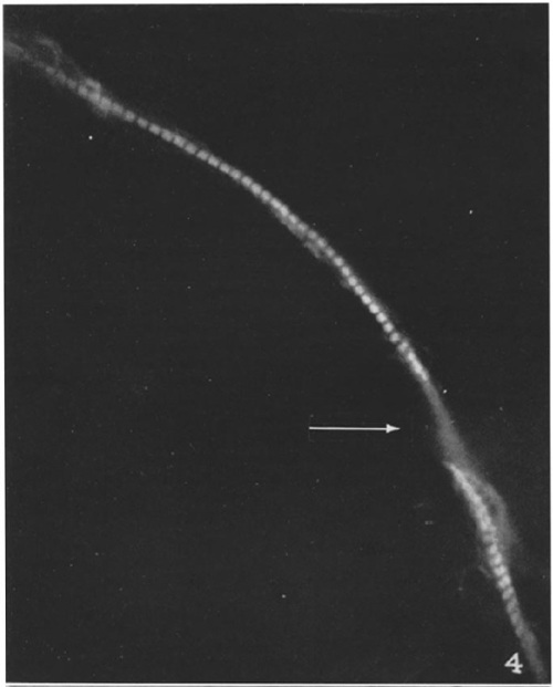
A single myoblast isolated from a stage 23 chick embryo, stained with rabbit globulin specific for myosin (1100x). The arrow indicates where the nucleus obscures the myosin. (Holtzer et al., J Biophys Biochem Cytol 1957)

glomeruli-ortega-1956

Kidney of rat injected with nephrotoxic rabbit antibodies to cause nephritis; the rabbit antibodies concentrate in the glomeruli, as seen by staining with goat globulin specific for rabbit globulin (37x and 205x). (Ortega and Mellors, J Exp Med 1956)

Pubmedwhack: Coproagglutinins

You may be familiar with the concept of a “Googlewhack”, which in common parlance is a word or phrase that returns exactly one result when typed into Google. Wikipedia claims it must be two words, without quotation marks, and a single word or a phrase in quotation marks is not a “Googlewhack” but is a “Googlewhackblatt” or some other hair-splitting neologism. But the word was invented and given that definition a decade ago, and I think the original definition is no longer practical.

So a Pubmedwhack would be a phrase that, when typed into Pubmed, the indispensible bioscience citation database, gives exactly one result. Like “vampyric” (1), or “great blue heron embryos” (2).

One example from immunology is “coproagglutinins”. Search for that and all you get is a 1951 paper (3) (subscription required) from the U.S. Army’s 406th Medical General Laboratory, which was headquartered in Tokyo.

coproagglutinins

In this study they were trying to diagnose which type of organism was causing bloody diarrhea, by looking for antibodies in the diarrhea itself. Anti-Shigella antibodies suggest that it’s a Shigella infection. Anti-Corynebacterium antibodies suggest diphtheria. And so on. “Copro-” is, of course, the prefix referring to feces, as in “coprolite” (4). The word “coproantibodies” is found in a few dozen titles and abstracts, including 20 or so from the 21st century. So it’s not a ridiculous technique. But this is the only paper to combine the new (as of 1951) concept of antibodies measured in feces, with the old (as of a few years later) word “agglutinins”.

To detect antibodies at this time, we had moved beyond complement fixation and amboceptors and whatnot. People doing serological tests could mix serum, or in this case fecal extracts, with a sample of Shigella paradysenteriae. If the extract contained antibodies specific for that bacteria, they would bind each other and form aggregates, making the solution cloudy.

That’s agglutination. You do it to detect agglutinins, which are the kind of antibody that agglutinates. Although agglutinins, like amboceptors, are best described as simply “antibodies”.

Now we have various other techniques for detecting antibodies. So to call them “agglutinins” just because you’re using agglutination to detect them adds confusion.

The word “agglutinins” is still used in the 21st century, to describe the antibodies of certain autoimmune diseases, as in this Blood editorial (5). We all make some antibodies against our own red blood cells. With too many anti-RBC antibodies, this can lead to the RBCs aggregating and being destroyed by complement. Especially in cold weather and in the extremities, hence the name “cold agglutinins”.

Science is not aware of any cold coproagglutinins.

Another way to tell Salmonella from Shigella (click for source)

Another way to tell Salmonella from Shigella (click for source)

1. Almond BR (2007). Monstrous infants and vampyric mothers in Bram Stoker’s “Dracula”. Int J Psychoanal 88:219-235.

2. Hart LE (1991). Dioxin contamination and growth and development in great blue heron embryos. J Toxicol Environ Health 32:331-344.

3. Barksdale WL, Ghoda A, Okabe K (1951). Coproagglutinins in ulcerative colitis. J Inf Dis 89:47-51.

4. Steve (2010). Coprolites: A few words on prehistoric turds. WebEcoist.Momtastic.com, accessed November 1, 2013.

5. Stone MJ (2008). Heating up cold agglutinins. Blood 116:3119-3120.

Rabbit mosaic virus?

This paper (1) seemed weird, at first. With a title like “Immunologic Reactions with Tobacco Mosaic Virus“, and methods that included injecting the virus into rabbits, I wondered if it was an attempt to use all possible means to infect animals with the legendary plant virus. But no, the author of today’s paper points out in her introduction that that work was already done (2) by Maurice Mulvania of the Tennessee Experiment Station, and we now know (as of 1929) that the virus doesn’t reproduce in rabbits.

So what is she doing, looking at the rabbit’s immunologic reactions to tobacco mosaic virus (TMV)?

Well, primarily, trying to back up the evidence that the virus exists at all, as a physical object. The introduction to this paper is particularly interesting in its snapshot of what people thought about viruses, before electron microscopy.

The properties of tobacco virus are frequently linked to those of enzymes. An apparent similarity between them may be due to the fact that they have both been studied in tissue extracts and in an impure state.

In Angela Creager’s 2001 book The Life of a Virus: Tobacco Mosaic Virus as an Experimental Model, 1930-1965, a similar point is made:

Investigations of virus infectivity resembled contemporary biochemical studies of enzyme activity under various conditions. Allard, mentioned above for his insistence on the contagious nature of TMV, assessed the effects of heat, germicidal agents, and chemical reagents on viral infectivity… A few years later, Maurice Mulvania conducted similar studies assessing the effects of light rays, heat, dialysis, and exposure to bacteria on the infectivity of TMV, publishing his work in the other major journal for research on plant diseases, Phytopathology. The susceptibility of TMV to some of these inactivating agents led him to speculate that the virus might be a simple protein or enzyme.

So the word “virus” did not mean “virus” as we now know it, an organism with genetic material that can reproduce when it infects host cells. A “virus” could simply be a toxic substance. After all, “virus” comes from the Latin word for poison.

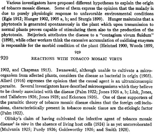
Helen Purdy, later Helen Purdy Beale, was a legendary figure in plant virology. Here is a 2006 review of her work (3) that mentions, right off the bat, this 1929 paper in which she immunized rabbits against TMV-infected plant saps and uninfected plant saps, and concluded that the virus saps had unique proteins which were not present in the normal plants. In the introduction she also gives a mini-literature review of what people thought TMV might be.

purdy-1929-introduction

Verdict of history:

Light and electron microscopy of cauliflower mosaic virus in Brassica rapa cells, from Martelli and Castellano, 1971 (5). "IB" means inclusion bodies.

Light and electron microscopy of cauliflower mosaic virus in cells, from Martelli and Castellano, 1971 (5). “Ib” means inclusion bodies. Click for higher resolution.

  • Egiz: Wrong!
  • Hunger: Wrong! That sounds like a prion disease.
  • Beijerinck: Right about the “contagium vivum” part, wrong about “fluidum”. Beijerinck is generally credited as the first scientist to demonstrate that an infection could be transmitted by something smaller than a bacterium.
  • Heintzel/Woods/Chapman: Wrong!
  • Iwanowski: Right about being unculturable, wrong about being bacterial.
  • Allard: Right!
  • Palm: Right! Those inclusions are made up of solid particles of the infectious virus.
  • Olitsky: Wrong!

Even Palm didn’t contemplate a new form of “parasite” other than extra-tiny bacteria. His paper (4) was written in Dutch and published in the now-elusive Bull. Deliproefsta. te Medan-Sumatra. This publication is so elusive that I can’t figure out what “Deliproefsta” stands for. Among others interested in Palm’s findings, Dr. L.O. Kunkel provided a synopsis at the end of an article in the 1921 Bulletin of the Experiment Station of the Hawaiian Sugar Planters’ Association. (This is what came up first from a web search for “tobacco” and “deliproefsta”.

In a recent paper Palm has described certain “amoebiform corpuscles” which he finds in mosaic tobacco leaves. He apparently considers these bodies to be analogous to those of corn mosaic. In addition to the “larger foreign corpuscles,” which he says “lie either in intimate contact with the nucleus or more or less in its vicinity”, he finds numerous extraordinarily small granules which, he thinks, may represent an orgnaism. On the assumption that the granules are alive and that they are the same bodies which Iwanowski described as bacteria, he gives them the name Strongloplasma iwanowskii.

In considering the intracellular bodies as a possible cause of mosaic disease, the writer meets the same difficulty which prevented Iwanowski from looking on the “Plasmaanhaufungen” as causal organisms. They are too large to pass through bacterial filters. Iwanowski did not know, however, that these bodies are associated with mosiac disease in several different plants. Perhaps this further evidence may justify an attempt to harmonize statements which at first appear to be contradictory. It may be that the amoeboid bodies… represent only one stage in the life of a causal organism. At another stage they may be so small and plastic that they can pass through the fine pores of a filter and escape detection under the microscope. They probably become visible only after a certain period of growth within the host cell.

Bingo!

* * *

The 1929 Purdy paper has a remarkably coherent set of conclusions that agree with what we now know. Purdy did a large number of experiments that mostly involved immunizing rabbits with “virus-saps” and “normal saps” from tobacco, tomato, pepper and petunia plants. She then took the immune sera from these rabbits and saw if they could block the activity of the various saps. There’s a whole page of “summary” and conclusions. In brief:

  • Tobacco virus-saps contain all the antigens that normal tobacco saps contain, plus a few others.
  • Virus-saps from tobacco, tomato, pepper and petunia plants all have some antigens in common, which are probably not found in normal sap.
  • Normal rabbit or guinea pig serum cannot block TMV from infecting plants. Neither can serum from animals immunized with normal plant sap. These sera do not contain the proper antibodies. But serum from animals immunized with virus-saps can block TMV from causing plant disease.

Well, that’s pretty conclusive. But these plant saps are mostly just soluble proteins. So we still don’t know if the virus is made of of particles, or if it is liquid in nature. Some sort of extra-sensitive microscope would be very helpful.

* * *

1. Purdy HA (1929). Immunologic reactions with tobacco mosaic virus. J Exp Med 49(6): 919-935.

2. Mulvania M (1926). Studies on the nature of the virus of tobacco mosaic. Phytopathology XVI: 853-872.
(note: I can’t find any archive that includes Phytopathology any earlier than 1970, which is a shame since a lot of papers and indices refer to Mulvania’s work).

3. Scholthof KBG, Peterson PD (2006). The role of Helen Purdy Beale in the early development of plant serology and virology. Adv Applied Microbiol 59: 221-241.

4. Palm BT (1922). Die Mosaikziekte vac de Tabakeen chlamydozoonose? Bull Deliproefsta te Medan-Sumatra xv: 7-10.

5. Martinelli GT, Castellano MA (1971). Light and electron microscopy of the intracellular inclusions of cauliflower mosaic virus. J Gen Virol 13: 133-140.