Amboceptor

Microbiological, virological, bacteriological, immunological, medical, epidemiological, historical, anecdotal

Category: Virological

This week in bad ideas: Wart grafting

good-times-warts

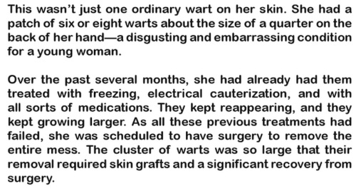
As described in this excerpt from James G. McCully’s Good Times In The Hospital, warts can be so extensive that they require a skin graft.

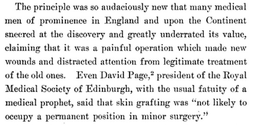
In the 19th century, skin grafts took many forms. By 1912 the process was down to a science, as detailed in Leonard Freeman’s Skin Grafting for Surgeons and General Practitioners. The basic method was established, with individual variations (“Donnelly, for some unexplained reason, prefers grafts from a portion of the skin subject to slight motion, such as the insertion of the deltoid muscle”), and complications and shortcomings were well known. (“Of all constitutional disorders, syphilis is probably the most disturbing, so much so that it has been claimed that grafting should never be attempted when this disease is present, and Freeman cites several cases which he thinks show that grafts will not adhere until syphilis has disappeared from the system.”) Many advancements had recently been made, even surprising doctors who didn’t quite expect them to work, and didn’t have a real theory to explain their success. The words of the late Dr. David Page (quoted here in 1872) were recalled with pity.

david-page-fatuity

Freeman’s book, like other contemporaneous reviews of skin grafting techniques, includes an extensive section on “anomalies in skin grafting”. This included inducing blisters and transplanting the thin blister roof; transplanting hairs (taking care to include the hair sheath); skin from a wide range of different animals, particularly frogs; bits of muscle; egg membrane; and of course, thin sections of rabbit testes.

E. Aievoli made use of thin sections of the testes of rabbits for purposes of grafting in four cases, assuming that the testicle possesses a greater cellular activity than other portions of the body. The results were undoubtedly good, but it does not follow that they were better than could otherwise have been obtained.

It’s obvious now that most of these substances would not fuse with, or become, actual skin, but they provided a suitable covering for the wound / ulcer / burn until it could heal. From a 1909 review in the Boston Medical & Surgical Journal, by Dr. Albert Ehrenfried:

Orcel (1888) states his belief that animal grafts act merely as a sort of protective dressing, and he is substantiated by the experiments of Beresowsky (1892).

Ehrenfried has a particular interest in converting surgeons to the “Reverdin method”, which sounds simple, safe, and advantageous in that the source of the graft is easy to obtain. You just take “morsels” of existing skin, aiming for a maximum of 1/8 inch in diameter (but cut from as deep as possible), and “seed” them over the “granulations of ulcers”. Held in place with “diachylon plaster”, they should allow healing to proceed faster, as it will spread from these new pieces of skin as well as from the margins of the ulcer.

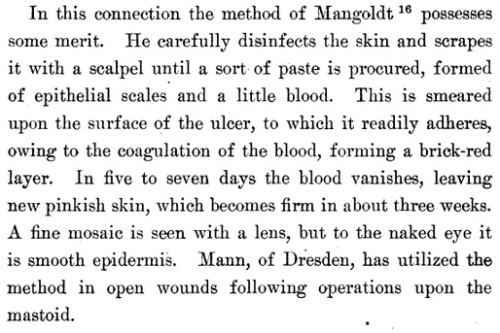
Reverdin preferred this to the even easier “epithelial dust” method of Mangoldt.

method-of-mangoldt

And another procedure, that might make more sense than the early 20th century writers think, is sponge grafting, “introduced by Hamilton in 1881.”

Sponge does not grow fast to the surface as does skin, but acts merely as a stimulating support for the granulations, finally undergoing complete absorption. The procedure is much inferior in its results to the transplantation of cuticle, and is now seldom employed.

A fine Turkey sponge is selected, soaked in dilute nitrohydrochloric acid until all calcareous particles have been dissolved, and then placed for a time in desired in a solution of potassium hydroxid. Very thin slices, which are the most serviceable, can be cut … and sterilized by boiling, or in a 5 per cent solution of carbolic acid.

The sponge is then spread upon the granulations, which have been rendered as nearly aseptic as possible (see the method of Reverdin), and dressed much as if it were a transplantation of skin. The granulations soon acquire new energy and push their way into the interstices of the sponge, which often almost disappears beneath them, so luxuriant is the growth.

Providing an aseptic framework for tissue to grow around is something bioengineers do now, right? Albeit not with skin tissue.

* * *

But there’s one skin-grafting method that might have extra negative effects, beyond introducing infection, irritation, or just wasting everyone’s time. At the top of this piece, we see how skin grafts may be necessary for a bad case of warts. But what if you have a bad case of burns, from molten iron spilling into your boot? You also need a skin graft… and as a source of the new skin, you could use warts!

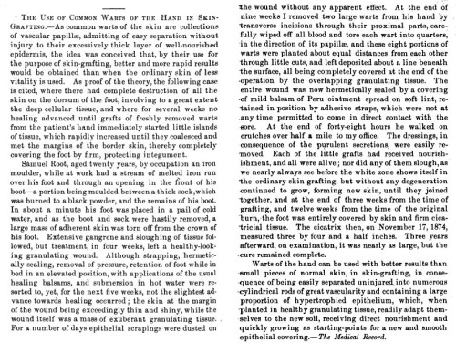
The first (and only?) example of this procedure was described by Dr. Charles Leale of New York in an 1878 report in The Medical Record. Presumably this was the same Charles Leale who 12 years earlier had attended at the Lincoln assassination, as a 23-year-old army doctor who would soon leave the public eye for many decades of lucrative obscurity. (Obscure enough that in some of the journals that reprinted his observations on wart grafting, he was credited as Charles Seale or Charles Lale.)

The Medical Record seems not to be archived in Google Books. The most comprehensive summary of Leale’s “The Use of Common Warts of the Hand in Skin-Grafting” now available is probably in the September 1878 Atlanta Medical and Surgical Journal, vol. XVI(6):354-355. Here’s an abstract, and then the whole thing. Why not, it’s only two pages.

As common warts of the skin are collections of vascular papillae, admitting of easy separation without injury to their excessively thick layer of well-nourished epidermis, the idea was conceived that, by their use for the purpose of skin-grafting, better and more rapid results would be obtained than when the ordinary skin of less vitality is used. As proof of  the theory, the following case is cited, where there had complete destruction of all the skin on the dorsum of the foot, involving to a great extent the deep cellular tissue, and where for several weeks no healing advanced until grafts of freshly removed warts from the patient’s hand immediately started little islands of tissue, which rapidly increased until they coalesced and met the margins of the border skin, thereby completely covering the foot by firm, protecting integument.

leale-warts-entire

Do we know if it’s possible to transplant warts and have them take root in new places? If it didn’t happen in this case, as Leale says it didn’t, it may be very hard to do.



Data update: Destroyed by freezing does not equal alive

In the experiments reported in the present paper a number of active agents, some undoubtedly living, others equally unquestionably not living, and still others of a doubtful nature, were subjected to repeated freezing (-185°C.) and thawing. By these tests it has been possible to determine that mere destruction or inactivation of a substance cannot be accepted as proof that it existed in a living state.

In 1926, legendary virologist / bacteriologist Thomas M. Rivers addressed the still-extant question “Are viruses alive?” through the simple experimental method of freezing them over and over. The paper in question came out in the Rockefeller Institute house publication Journal of Experimental Medicine (vol.45[1]: pp. 11-21). Not exactly a gripping title.

repeated-freezing-rivers

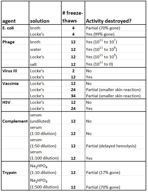
What kind of data is in here? Well, for most of the graphs they take an “active agent” (either bacteria, virus, or enzyme), freeze-thaw it “as often as desired”, and then see if it’s still active. The data is pretty straightforward, with a separate section for colon bacilli, a section for Virus III, a section for “a bacteriophage lytic in colon bacilli”, and so on. But it’s never summarized in a way that compares the different “agents” to each other. Let’s try to do that.

The low temperature (-185°C.) used in the experiments to be reported was obtained by means of commercial liquid air which was transported from the plant to the laboratory in Dewar flasks. Desired amounts of the air were transferred to deep Dewar beakers where small amounts of the substances to be frozen, enclosed in Noguchi tubes, were completely immersed for several minutes. After the substances had been completely frozen they were quickly thawed in tap water (16-18°C.).

What does “active” mean for these various substances? How did they determine that freeze-thawing had destroyed the substance’s activity? This was different for each substance.

rivers-activity

Bacteria can be measured the same way we measure them now, by making serial dilutions and plating each dilution on agar, then seeing how many colonies grow. Bacteriophage can be measured the same way, by first making a “lawn” (agar plate fully covered with bacteria) and applying different dilutions of bacteriophage, then seeing how many “plaques” (holes in the lawn) are formed by the phage killing the bacteria.

Complement can be measured by seeing how long it takes to destroy “given amounts of red blood cells in the presence of a great deal of amboceptor“.

Trypsin can be measured somehow, Dr. Rivers doesn’t specify except by saying that his colleague Dr. Northrop took care of that part of the study. Nowadays Dr. Northrop would be a co-author. But the paper would then have to list Rivers as both the first author and the last author somehow, since Northrop didn’t do enough to merit either status.

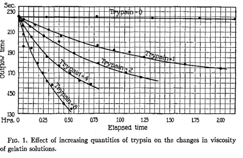
Anyway, to find “details of the technic” we are directed to a paper by Northrop and Hussey (1923), showing a very clever method by which a solution of gelatin is exposed to trypsin, and at different timepoints the gelatin’s viscosity is measured.

northrop-graph

And how do you measure viscosity? With a viscosimeter.

northrop-viscosimeter

I’m having trouble understanding sentences like “The gelatin-water time ratio was approximately 3”, but the point is that you can measure the amount of trypsin by measuring how fast it turns gelatin into runny gelatin. Nowadays you would use a colorimetric assay, in which trypsin cuts the protein that has some sort of colored label attached to it, and you would measure how much colored label gets released into solution.

* * *

Finally, the three mammal viruses.

“Vaccine virus” and “Virus III” are both introduced to the skin of rabbits, probably by scarifying and then rubbing the virus into the scratches. They look for a “virus reaction” in the skin surrounding where the virus was inoculated, and measure this semi-quantitatively based on how bad of a sore forms. I hope they aren’t simply measuring the immune response to the inoculation, because even killed virus should produce some immune response.

“Vaccine virus” is basically what we now call vaccinia virus. “Virus III” is a more interesting question.

All stocks of Virus III were lost some time before the invention of electron microscopy. Nobody can now be sure what exactly this rabbit-specific virus was, but it was probably Leporid herpesvirus 2, as described by Nesburn in 1969. For an objective summary of the Virus III story, read the page on LHV-2 (p. 380) in The Laboratory Rabbit, Guinea Pig, Hamster, and Other Rodents (Academic Press, 2012).

“Herpes virus” (Herpes simplex) activity is measured differently. They inject HSV into rabbit brains, and they look for dead rabbits. It seems like this assay should actually be quantitative, based on looking at how long it takes the rabbits to die, but all their rabbits either died within a week or lived longer than a month, so the results were clear without measuring time to death.

 * * *

The results are pretty simple, which is why I would like a summary figure instead of a series of tiny individual tables for each thing they studied.

rivers-summary

Some comments on the data:

  • “Locke’s solution” is like what we call Ringer’s solution, the intravenous fluid given to people who have suffered blood loss. This page indicates that it’s the same as Ringer’s solution, but buffered by bicarbonate instead of lactate. But most other sources indicate that it also includes glucose, making it closer to Tyrode’s solution.
  • Locke’s solution is basically a physiological salt solution, so what’s the difference between that and the “physiological salt solution” used in the phage experiments? The latter doesn’t contain glucose, and isn’t buffered; instead of a pH of roughly 7.6, the pH is “between 6 and 7”.
  • Vaccinia virus was the most stable, retaining the ability to create a skin lesion even after 34 freeze-thaws. Virus III was destroyed after 12, and the other herpesvirus was destroyed after 24.
  • Both bacteria and bacteriophage had more than 99% of their activity destroyed by freeze-thawing in Locke’s solution. All three big viruses were more hardy than that.
  • I don’t know what a “1:10 dilution of trypsin” is. How many USP Trypsin Units is that? What’s the molarity? We have to take Dr. Northrop’s word for it, that it’s the same system for trypsin dilution he always uses.
  • At a mere 1:10 dilution, complement and trypsin were not damaged by 12 freeze-thaws. But when diluted further, they were very susceptible to freeze-thaw.
  • In fact, bacteria and bacteriophage were also more susceptible to freeze-thaw at low dilutions. Even vaccinia virus lost all its effect after 34 dilutions at 1:100,000 dilution, although it didn’t have much activity at that dilution to begin with.
  • Of further interest: the difference between high and low dilutions was only observed in Locke’s solution or salt solution, not broth. Remember these cutting-edge findings when you make your own aliquots.

Viruses can be RE-activated by light?

When you’re always looking at old sources, you run the risk of condescending to the experts of the past, who believed scientifically plausible things that now seem obviously wrong. More than once I’ve been ready to point out some amusing practice of the distant past, only to find out that it’s a perfectly valid fact that I (having no medical or physiological training) had never heard of.

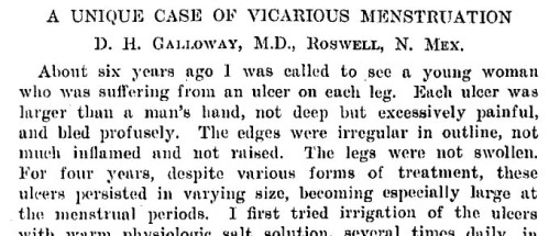
One example is “vicarious menstruation”. Is it possible that menstruation could manifest as a nosebleed, or sores in the mouth (sometimes called “herpes”)? Or as a pair of ulcers on the legs, as D.H. Galloway of Roswell, New Mexico reported in 1913? Isn’t it more likely that these stories are exaggerated, or are coincidental? But yes, some combination of hormone levels and blood pressure creates that phenomenon in some women.

galloway-vicarious-menstruation

Another one is “activated milk”, which contained a substance called “viosterol” that was in high demand for preventing rickets in children. Activated milk? Activated by what?

Ultraviolet light, it turns out. Did this work? Well, UV light turns cholesterol into vitamin D3 when our own bodies are exposed to the sun, and it turns the fungal (yeast) equivalent, ergosterol, into vitamin D2. Cows could be fed UV-activated yeast to make them produce “activated milk”, or activated yeast extract could be directly added to milk. Either of these was a way of “activating” milk that probably worked. Exposure of normal milk to UV light seems like it would be a waste of time.

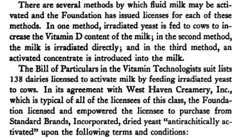
Whether its benefits were exaggerated or not, activation of milk and other foods was extremely popular, as described in Michael Holick’s great historical review in Public Health Reports, called “The Vitamin D Deficiency Pandemic: a Forgotten Hormone Important for Health”. The drug and food industries fought over whether companies like Fleischmann’s Yeast could claim their products were the equivalent of vitamin D supplements. Here’s a contemporary excerpt from Cartels: Challenge to a Free World, Wendell Berge’s 1944 classic of vaguely paranoid economics.

cartels-activated-milk

And all the way into the 21st century, there’s heated debate over whether vitamin D2 (the vitamin D in most supplements) is an appropriate substitute for our own vitamin D3.

* * *

So anyway, here’s another real thing that looked weird and debunkable at first glance.

In virology papers from the fifties and sixties, there are many mentions of something called “photoreactivation”. This started with 1949 work by future Nobel laureate Renato Dulbecco, done in the Indiana University laboratory of future Nobel and National Book Award laureate Salvador Luria. In 1950 Dulbecco summarized the story.

Kelner (1949), working with conidia of Streptomyces griseus, discovered that light belonging to the visible range is capable of reactivating biological material that has been rendered inactive by ultraviolet radiation (UV). Shortly after Kelner’s discovery was known, a similar phenomenon in bacteriophages (bacterial viruses) was observed by accident. Plates of nutrient agar containing UV-inactivated phage and sensitive bacteria had been left for several hours on a table illuminated by a fluorescent lamp. After incubation it was noticed that the number of plaques was higher on these plates than on similar plates incubated in darkness. A short report of this phenomenon of “photoreactivation” (PHTR) has already been published (Dulbecco, 1950).

We’ve been using UV light, gamma rays, and chemical agents like nitrogen mustard to make “killed” versions of viruses, safe for use in vaccines. And now it’s possible that visible light could then re-activate these menaces? Should vaccines be stored in the dark?

Beyond  bacteriophages, many other viruses were found to be capable of photoreativation. A sample:

  • 1955: “Of the three viruses we studied earlier, tomato bushy stunt and the Rothamsted tobacco necrosis virus showed the phenomenon of photoreactivation, and tobacco mosaic virus did not … Of the six viruses that did [in this study], potato X showed it much the most strongly, tomato bushy stunt and a tobacco necrosis virus the least; cabbage black ringspot, cucumber mosaic and tobacco ringspot were intermediate.”
  • 1958: “Thirty minutes of illumination at 300-380 f.c. gave substantial photo-reactivation [of] potato virus X”
  • 1961: Tobacco mosaic virus particles can’t be photoreactivated, but RNA preparations from the virus can.
  • 1967: “Photoreactivation of UV-irradiated blue-green algal virus LPP-1”
  • 1967: “By contrast, photoreactivation of the irradiated [tobacco necrosis virus] was observed in French bean and tobacco, but not in Chenopodium.”
  • 1968: Pseudorabies virus can be photoreactivated in chick embryo cultures, but not in rabbit kidney cells.

In the last of those quotes, it’s becoming clear that viruses don’t photoreactivate on their own. They photoreactivate inside cells. You can use UV light to damage the DNA (or RNA) of a virus so it can’t multiply. But it may still infect cells if the protein coat is intact. Then once the viral DNA (or RNA) is inside the cell, the cell’s DNA repair mechanisms can go to work. One of these is photolyase, found in plants, bacteria, fungi, and some animals, but not mammals. Blue light activates this enzyme to reverse the DNA damage caused by UV light (specifically, covalently-linked pyrimidine dimers).

So instead of thinking of photoreactivation as something that happens to certain viruses, we should think of it as something that happens in certain types of cells, to viral DNA as well as cellular DNA.

By 1958, Dr. John Jagger (who does not have a Wikipedia page, though his wife, also a scientist, does) was already able to write a fantastic review of photoreactivation in general (not just viruses and bacteria), saying:

Photoreactivation seems to be possible whether the UV damage occurs in the liquid or the solid state. However, the reactivation seems to require not only the liquid state, but a rather complex environment, similar to that within a living cell.

It doesn’t quite require a living cell, but it requires “cellular material”. A cellular extract still contains the photolyase enzyme.

* * *

You’ll notice that the above examples are almost all plant viruses. This is partially because plants were a very convenient system for virology in the era before cell lines, but it also has to do with the importance of light in plant biology. Dependent on the sun, they need to be able to counteract the negative aspects of ultraviolet light.

But it’s also clear that photoreactivation takes place in insects and fish.

The data show that fish cells have an efficient photoreactivation system at wavelength > 304 nm that reverses cytotoxicity and dimer formation after exposure to filtered sunlamp irradiation of a shorter wavelength (lambda > 290 nm). Shorter wavelengths in UVB (> 304 nm) are more effective in photoreversal than longer ones (> 320 nm). As a consequence, 50-85% of dimers induced by these wavelengths in fish are photoreactivated while they are being formed. A major cytotoxicological lesion is the cyclobutane pyrimidine dimers. Cultured human fibroblasts do not possess such a repair system.

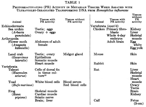
What about that paper above, in which chicken embryo cells enable pseudorabies virus (a herpesvirus) to reactivate? That looks weird, to me at least. Shouldn’t chickens, being warm-blooded animals, be grouped with mammals rather than fish? But chickens aren’t mammals. This table, from Photoreactivating-enzyme activity in metazoa [Cook JS, McGrath JR [1967] PNAS 58(4):1359-1365] sums it up.

photoreactivation-in-metazoa

Mammals have other DNA repair mechanisms, but we lack photolyase. Which as it turns out, makes us kind of weird.

Yellow fever stops at the Miami airport.

staining-mosquitoes-before-liberating

Initially, new infectious diseases could spread only as fast and far as people could walk. Then as fast and far as horses could gallop and ships could sail. With the advent of truly global travel, the last five centuries have seen more new diseases than ever before become potential pandemics. The current reach, volume and speed of travel are unprecedented, so that human mobility has increased in high-income countries by over 1000-fold since 1800. Aviation, in particular, has expanded rapidly as the world economy has grown, though worries about its potential for spreading disease began with the advent of commercial aviation. [1]

Podcasts are great. But in the world of science podcasts, many are simply boring because there is only one person talking, or one person interviewing another person. Unless it’s slickly produced and edited (the Nature or Science Times podcasts), I’ll quickly lose interest.

It’s better when the podcast is three or four people who know each other, having a conversation. This is is the format of the TWi[X] series, particularly the flagship This Week in Virology. TWiV co-host Dickson Despommier, though not a virologist, contributes a big-picture point of view when the show leaves its territory of basic lab science and moves into epidemiology and patterns of disease outbreaks. Here’s his lecture on how West Nile arrived in North America and spread from state to state, energized by a very hot summer at the Bronx Zoo.

More than once I recall Dr. Despommier pointing out that though it’s all well and good to model how a epidemic might spread by looking at the day-to-day movements of people and mosquitoes, the most powerful and mobile disease vector is… the airplane.

That fact has become ever more clear with the SARS and West Nile outbreaks, as we used genetic analysis to track them across the continents in real time. But you might be surprised at how long ago people recognized the airplane as a challenge to disease control.

* * *

80 years ago, yellow fever was the archetypal mosquito-transmitted disease. Particularly as the disease was endemic in some parts of the world (e.g. South America), but not in others (e.g. North America), despite both places being home to the vector, Aedes genus mosquitoes. A 1934 article [2] coauthored by Henry Hanson, veteran of battles against yellow fever on three continents, points out that dengue fever (also transmitted by Aedes) had reappeared in Florida after an eleven-year absence, and yellow fever could do the same at any time.

dengue-1922-1934-tampa-miami

Hanson and coauthor T. H. D. Griffitts go on to chide the cities of Florida and Georgia for their complacency in providing countless unsupervised water vessels in which the mosquitoes can reproduce.

Practically every urban community in the South has its array of artificial containers, from flower vases to catch basins, cuspidors and discarded automobile tires, producing Aedes aegypti. For example, the city of Tampa in eight weeks reported finding 1,091,823 containers (potential mosquito “hatcheries”). Of these, larvae were found in 20,864, or approximately 2 per cent. This was an unusually dry season (average rainfall of 1 1/2 inches a month for this period). It is interesting to note that in Miami for a like period only 56,598 potential breeding containers were reported, with 38,401, or 68 per cent, of the total actually breeding.

So I made an error above. The vessels aren’t quite “countless”. There are 1,091,823 of them in Tampa, give or take a dozen.

Though concerned that yellow fever could reach North America (again), Hanson and Griffitts are more concerned about India. The disease is only known in jungle areas of Brazil, Colombia and Bolivia (they claim); by comparison, Africa is its ancestral “home” and India, fairly nearby by plane, is virgin territory for yellow fever epidemics.

Today there is a feeling of concern … that Old World territory, where the vectors abound and where yellow fever has never before stalked, may experience widespread and devastating epidemics. One infective mosquito traveling in an airplane from the home of yellow fever (Africa) to India could be the spark to start the conflagration.

Thus there was considerable surveillance for mosquitoes and infected passengers at airports worldwide.

The earliest article I can find specifically discussing airplanes as disease vectors is from 1930, the first [3] of two identically titled 1930s editorials in the American Journal of Public Health. I don’t know if the international rules discussed here were enacted, but they show awareness that quarantine and disinsectization measures developed for ocean travel need to be multiplied and intensified to cope with passenger aircraft. Click for a bigger view.

airplane-and-yellow-fever-1930

* * *

For the purpose of determining whether or not mosquitoes are carried in airplanes, and, if so, to what extent, the distance of such transportation, the species of mosquitoes, and the type of planes on which they are carried, the United States Public Health Service began, on July 23, 1931, the inspection of all airplanes from tropical ports arriving at the airports of the Pan American Airways System at Miami. [4]

An anecdote from less than a year later illustrates how the search for mosquitoes had become a normal part of a plane’s arrival in the U.S. from South America.

A very Normal Rockwell scene of mosquito inspection. From LIFE Magazine, 5/27/40.

A very Normal Rockwell scene of mosquito inspection. From LIFE Magazine, 5/27/40.

The story of “the first international aerial hitchhiker” is as follows.

Paul Kaiser, 25, tried to immigrate from Czechoslovakia to America via a circuitous route. First, he got to the German port of Bremen, from which he sailed to Colón, Panama on an ocean liner. Sneaking into the nearby Canal Zone airport, he climbed into the baggage compartment of a “big 22-passenger Commodore plane”. The plane first landed in Barranquilla, Colombia, where he was not detected. It then landed in Kingston, Jamaica, where he was not detected. Finally, after Kaiser had spent 2 days without food, he landed in Miami. However, in Miami “a very thorough inspection is given every plane for mosquitoes, for there is always danger of the deadly yellow fever mosquito surviving the short trip and landing in the United States, a most undesirable immigrant”. Check out the April 1933 issue of Flying magazine for more details.

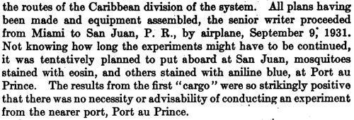
A few months earlier, T.H.D. Griffitts (coauthor of the aforementioned Henry Hanson article) performed the first experiments on mosquito transport by aeroplane. These were published in late 1931, in an enjoyably conversational article in Public Health Reports [4].

Dr. Griffitts, you don't really have to describe the experiments you WANTED to do but then decided were unnecessary.

Dr. Griffitts, you don’t really have to describe the experiments you WANTED to do but then decided were unnecessary.

T.H.D. Griffitts (and J.J. Griffitts — his son?) start out by describing all the mosquitoes observed on normal commercial flights between July and September of 1931. Most were Culex quinquefasciatus, but several other species were observed including Aedes aegypti. In fact, a later Griffitts paper [5] indicates that the historic first mosquito discovered on a Miami-bound plane was Aedes aegypti. (It’s somewhat confusing that the insect apparently arrived on a “ship from San Salvador, El Salvador”, but I think that at this time the word “ship” could be used for aircraft. And San Salvador is definitely not a port city.)

Griffitts also put mosquitoes on three planes departing from San Juan, Puerto Rico, and looked for them upon arrival in Miami. Today it takes 2.5 hours to fly from SJU to MIA, but in 1931 they stopped at three other airports along the way and the average travel time was over 10 hours. A total of 100 mosquitoes were labeled with eosin dye (to distinguish them from non-experimental and Miami-resident mosquitoes). 22 were observed upon arrival in Miami, after 1,250 miles of flying, opening of doors and hatches, loading and unloading of luggage, etc. It seems obvious that the insects can be imported… but these experiments prove it.

* * *

Eight years after 1930’s “The Airplane and Yellow Fever” editorial, the American Journal of Public Health published another one [5], describing the policies for infectious mosquito control in Khartoum, Miami, and “the French Colonies and Mandates”.

As the 1930s progressed and India remained devoid of yellow fever, public health doctors kept amplifying their alarms about how devastating such an epidemic would be. From the 1938 editorial:

greatest-menace-in-the-world-today

75 years after that was published, yellow fever still hasn’t swept through India.

In 1938, the number of people who’d been vaccinated against yellow fever was less than a hundred thousand, mostly in Brazil. It had taken a long time to make a vaccine strain of YFV that was weak enough that it could be given as a live vaccine, but the 1938 trial (run by the Rockefeller Foundation) was successful, and in 1942 the number of people vaccinated was over 10 million. For his two decades of work on the vaccine, which along the way required multiple basic science breakthroughs for it to be manufactured in large quantities, Max Theiler of Rockefeller University received the 1951 Nobel Prize in Physiology or Medicine. Read more here [6] about Theiler’s story; read more here [7] about the many problems and hurdles that were overcome to end up with a safe vaccine. (I think the second one is open-access and the first one isn’t.)

Within a decade, though complications from the vaccine were common and the virus stayed endemic in the tropics, Theiler’s YFV vaccine had eliminated yellow fever as a source of large-scale epidemics.

* * *

Coda: As early as the airplane was recognized as a vector for disease… it was harnessed as a weapon against disease.

From a 1932 report in Science (8), Joseph Ginsburg of the New Jersey Agricultural Experiment Station explains how large regions of standing water that previously were inaccessible to mosquito control workers can now be reached by plane, so that the surface can be coated with larvicidal pyrethrum or oil.

cheesequake-killing-film


* * *

1. Tatem AJ, Rogers DJ, Hay SI (2006). Global transport networks and infectious disease spread. Adv Parasitol 62:293-343.

2. Griffitts THD, Hanson H (1934). Significance of an epidemic of dengue. JAMA 107(14):1107-1110.

3. Editorial (1930): The airplane and yellow fever. Am J Public Health 20(11):1221-1222.

4. Griffitts THD, Griffitts JJ (1931). Mosquitoes transported by airplanes: Staining method used in determining their importation. Public Health Rep0rts 46(47):2775-2782.

5. Editorial (1938): The airplane and yellow fever. Am J Public Health 28(9):1116-1118.

6. Norrby E (2007). Yellow fever and Max Theiler: The only Nobel Prize for a virus vaccine. J Exp Med 204(12):2779-2784.

7. Frierson JG (2010). The yellow fever vaccine: A history. Yale J Biol Med 83(2): 77-85.

8. Ginsberg JM (1932). Airplane oiling to control mosquitoes. Science 75(1951):542.