Amboceptor

Microbiological, virological, bacteriological, immunological, medical, epidemiological, historical, anecdotal

Category: Bacteriological

Data update: How dyestuffs make stuff die

churchman-parafuchsin

This year, antibiotic researcher Mark Wainwright published a “Discussion” in the International Journal of Antimicrobial Agents, suggesting that we should take another look at a subject that he works on but most scientists lost interest in 6 decades ago: highly toxic synthetic molecules that can be used as local antiseptics and have distinctive bright coloration. What he calls “photoantimicrobials” represent a subset (those that can be targeted more precisely, because they are activated by light) of therapeutic molecules that earlier generations simply called flavines, anilins, or “dyes”.

Wainwright tells us:

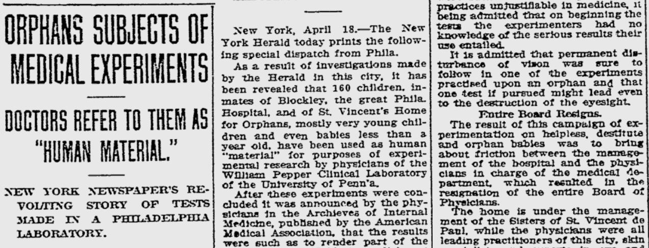
Browning introduced the application of the basic dyes named above [acriflavine, crystal violet and brilliant green] to battlefield wounds in 1914/15. The aim was always to achieve antisepsis and healthy wound closure, and the approach was also used in pre-operative sterilisation. The dyes used thus represent local antiseptics. Importantly, however, they were an improvement on the available hypochlorite solutions owing to the fact that they were not inactivated by fluids associated with wounds, such as serum. Fleming’s rebuttal of this work, noted above, was based purely on laboratory measurement. However, the disinfection of real wounds and the concomitant recovery of patients could hardly be argued against.

By far the majority of clinical work involving dyes was carried out before 1945. At this juncture, the penicillins and other antibiotics or natural product-derived antibacterial agents became the driving force in infectious diseases therapy. From the distance of the early 21st century, it is apparent that these wonder drugs have not been used correctly and that their overuse has allowed a much more rapid development of resistance mechanisms than might otherwise have been the case.

In terms of modern healthcare, dye therapy might be considered obsolete and ineffective, and this would indeed be the case if the performance criteria were still the same as in Browning’s day. However, recent research has developed a different approach, one that uses locally applied dyes in conjunction with light to provide a microbial killing effect. Such dyes may be referred to as photodyes or, more properly, photosensitisers. Given their remarkable utility in the field under discussion, they are commonly called photoantimicrobials.

The photoantimicrobial effect has been known in the laboratory since the turn of the last century, but has only been investigated realistically since the early 1990s and is currently proposed foruse in oral and ENT (ear, nose and throat) disinfection. Its remarkable activity lies in the in situ production of reactive oxygen species (ROS) on illumination. The highly reactive nature of species such as singlet oxygen, the hydroxyl radical and the superoxide anion ensures rapid oxidative damage to simple cells, sufficient to guarantee cell death. More importantly, there are no known microbial resistance mechanisms able to combat ROS produced in this way.

Furthermore, among the lead compounds used in photo antimicrobial discovery are the same dyes used by Ehrlich and Browning; methylene blue, crystal violet and acriflavine.

Brilliantly colored solutions for treating localized infections and washing out wounds may be making a comeback. And not just to treat pets, for which acriflavine, for example, is now used. In preparation for that retro craze, let’s look at some of the original pre-Depression research. churchman-gazette

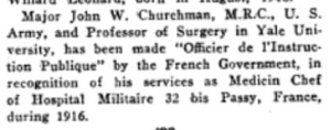
Most of it is in German, which I will ignore.

Much of the non-German work on “dye therapy” was led by urologic surgeon John W. Churchman of Yale University. At the top of this post is a rare color photo of bacteria in a Petri dish from the time when Julius Richard Petri (1852-1921) was still alive. This comes from a 1913 Journal of Experimental Medicine paper by Churchman, The Selective Bactericidal Actions of Stains Closely Allied to Gentian Violet, whose figures are otherwise black & white. Even the limitless moneybags of Old Elihu and the Rockefeller Institute only extended to printing one color figure.

* * *

Churchman’s papers are not very quantitative, but other “dye therapy” researchers did large-scale comparisons of multiple colored antibiotics, at multiple concentrations, against multiple bacterial species.

krumwiede-title

On January 1, 1914, the Journal of Experimental Medicine published the Observations of Josephine S. Pratt and Charles Krumwiede Jr. (not to be confused with their Further Observations, published 4 months later). They established a method for testing large numbers of drug/bacteria combinations while minimizing the amount of agar needed, by putting two samples in each Petri dish, slanted away from each other. Here they describe the process in vague terms that would be better presented as a 5-minute video.

As a routine a batch of agar was selected which was found especially suitable for the more feebly growing [bacteria]. To the hot agar an appropriate volume of a watery solution of dye was added to give the final dilution desired. The same agar was used in each experiment.

The most convenient and economical method was found to be as follows. One unopened Petri dish was used to tilt up one side of a second dish. In the opposite side was poured just sufficient agar to give a satisfactory slant. This dish was covered and used to tilt up a third dish, and so on in a row. After the agar had set, slants were poured in the other side of the Petri dishes which were tilted in the reverse direction. In this way two mixtures of agar could be used in the same dish, very little agar being required for each slant.

After inoculating all these plates with bacteria, they used the eye test. Compared to a control plate with no dyestuffs, how much did the dye prevent the bacteria from growing after 18 to 24 hours? Not at all? Was it “restrained”? “Markedly restrained”? Completely eliminated?

Here’s Table 1.

dyestuffs-original-table-1

That is a lot of information. 11 drugs, tested against 30 bacteria (12 Gram-positive and 18 Gram-negative), makes 330 combinations, of which only 7 potential tests were not performed. (I think the ellipsis means “not performed”.) Each of these 323 tests was done at 3 concentrations, making 969 data points in a single table.

To turn this into graphs would require many graphs. It would also require us to turn the X’s and +’s into a semiquantitative system (let’s say + = 4, ± = 3, X = 2, —* = 1, and — = 0). And since each data point is restricted to those 5 possibilities, you wouldn’t gain much by looking at a bunch of individual dots or bars anyway.

Let’s leave it as a table.

The main flaw of this table is that all the symbols look similar, except “—” . The bottom half of the table is a wall of symbols for different degrees of growth. It’s hard to see trends, because the symbols are hard to distinguish from each other, so it looks like every bacterium is resistant to every drug. Which is not quite true.

Instead of these text symbols, how about something that may be more intuitive — representing the 5 levels of growth as 5 colors. This may not be intuitive in every culture, and it may not work for the color-blind or in B&W printouts, but I took the liberty of changing the table. Now as bacteria proliferate, they pass from “no growth” (white) through yellow, green, and purple stages until they attain “growth like control” (charcoal grey). At first I wanted to range from white to black, but if the cells are black you couldn’t see the lines between them. Or maybe you can’t see them anyway.

For the tests that weren’t done, I replaced the ellipses with neutral grey. And finally, the two types of “diphtheroid bacilli” produced exactly the same results, so they were conflated into one.

dyestuffs-table-1

So where does this data lead us? Krumwiede and Pratt draw limited conclusions. The tables speak for themselves. To briefly summarize their Summary section: the “streptococcus-pneumococcus group” is more dye-resistant, and the “dysentery bacillus group” is highly unpredictable with dye-resistance showing “no correlation with the common differential characteristics”. Also, they discovered mutations that make bacteria lose resistance. (“Among Gram-negative bacteria a strain is occasionally encountered which will not grow on violet agar, differentiating it from other members of the same species or variety.”) Finally, in the middle of the Summary section they say this.

The reaction is quantitative, although the quantitative character is more marked with some species than with others.

Now, a modern reviewer would look unkindly on that sort of admission. First, it seems like it’s not quantitative, it’s semiquantitative. Instead of measuring the number of colonies, you’re grading “growth” on a scale from 1 to 5. And which are the species that don’t have a “quantitative character”? Why don’t you use some other method to see how well those ones are growing? And by the way, how repeatable is your eye test? And what does it mean exactly? Let’s see examples of “growth like control”, “restrained growth”, and “markedly restrained growth”. Does it mean there were fewer colonies, or the colonies were smaller, or both? And why use the same 3 concentrations of all 11 drugs? Maybe 1:500,000,000 would have still killed the bacteria, and be less toxic to patients.

That’s what I’d say, if the journal hadn’t told me “Your review is now 100 years late and consequently is no longer needed.”

* * *

One more paper: from 7 years later, Gay and Morrison in the January 1921 Journal of Infectious Diseases. Whereas Krumwiede & Pratt (1914) was the first in their series, this is a sequel.

gay-morrison-title

Krumwiede & Pratt took a limited range of dyes, and used then on every sort of bacteria they could find. Now Gay & Morrison use every dyestuff they can find on a limited range of bacteria. This is a much longer paper, so I’ll just address their first set of data, about bacterial growth in culture, and ignore their innovative rabbit empyema model.

Table 1:

gay-morrison-table-1

In this table, the numbers mean the reciprocal of the minimum inhibitory concentration (MIC). The MIC is the smallest concentration of the drug that will prevent bacteria from growing. If the MIC is 5 ng/ml, the bacteria should grow if the concentration is lower than that, and the bacteria should die if the concentration is 5 mg/ml or greater.

“2,000” means a 1 to 20,000 dilution. I think that’s weight/volume (1 gram in 20,000 milliliters). The lower the number in this graph, the more concentrated the drug needs to be to kill bacteria. A drug marked “2,000” needs to be a thousand times more concentrated to have the same effect as a drug marked “2,000,000”.

This table is clear. Intuitively we look at the numbers and recognize that 2,000 is smaller than 20,000 and therefore represents less of a dilution. If they were in scientific notation, it wouldn’t be as intuitive.

The only problem is the use of “0”. The most concentrated dyes they used were at a 1:2,000 concentration. If the bacteria still grew, they listed this drug as “0”, or not active against the bacteria. Taken literally, “0” means Staphylococcus would grow on media consisting entirely of methylene green. I would just write the word “inactive” instead of the number 0.

* * *

I might also expand the table to include the rest of the 40 dye molecules Gay & Morrison used in this study. That’s right — in addition to the 12 molecules listed above, they have data on Acid fuchsin, Acid violet, Acridine orange, Azo-acid red, Basic fuchsin, Benzo-azurin, Brilliant cresyl blue, Columbia blue R, Congo red, Crystal ponceau, Cyanin B, Diamil blue, Erioglaucin A, Janus dark blue B, Methylene blue (medical purity), Methylene blue GG, Neutral red, New fast green 3B, Nile blue, Oxamin violet, Rhodulin violet, Safranin, Sauer grün, Scarlet 6R, Setocyanin, Sulphon acid blue R, Toluidin blue, and Wasser blau.

But most of the data is in paragraph form. And it’s not immediately clear.

gay-morrison-excerpt

Let’s make an expanded version of the table above, containing all the dyestuffs. Even the dyestuffs that never killed any of the bacteria. And make some other changes:

  • Keep them arranged in order of effectiveness, but put the most effective ones at the top.
  • In fact, divide them into categories. Those that inhibited all 3 bacterial species, those that inhibited 2, those that inhibited 1, and those that were completely ineffective.
  • Update the nomenclature. We consider Bacillus typhosus to be Salmonella now.
  • And… why not color-code the chart. Maybe blue dyes will do one thing, and red dyes will do something else. Might as well include that information. Patterns may emerge.

gay-morrison-table-2

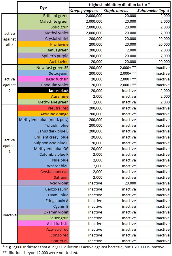
Isn’t it nice to see those colors? And it shows how much easier it is to kill Streptococcus pyogenes. There’s only one dye that kills either Staphylococcus or Salmonella but fails to kill S. pyogenes. And it shows that blue and red dyes aren’t very antibacterial, while green ones kill bugs dead. Why is that?

Wikipedia: Zinaida Ermolieva

Zinaida Vissarionovna Ermolieva (Russian: Зинаида Виссарионовна Ермольева; 15 October 1898 [O.S. 27 October] – 2 December 1974) was a Russian microbiologist and epidemiologist who led the Soviet effort to generate penicillin during the Second World War.


Biographyzinaida


Born on a farm in the Frolovo region, Ermolieva attended school in Novocherkassk and studied medicine at Don University in Rostov-on-Don (now part of Southern Federal University), graduating in 1921. Continuing to work at Don University’s bacteriological institute, she collaborated with Nina Kliueva on a study on encephalitis lethargica [1], before moving to Moscow in 1925. There she worked at the People’s Commissariat of Health, as head of microbiology at a biochemical institute [2] that would later be named for its founder Aleksey Nikolayevich Bakh [3]. Early in her career she was known for her work on characterizing lysozyme and employing it as an antimicrobial agent [4].

During the Second World War Ermolieva became famous for her role in the independent Soviet effort to extract penicillin from mold, using the species Penicillium crustosum [4] (rather than P. notatum, the species employed by Alexander Fleming and other British scientists). To test this penicillin treatment, she was one of many scientists to travel to Abkhazia and make use of the monkey colonies at Sukhumi’s Institute of Experimental Pathology and Therapy [5].

Ermolieva also led the efforts to control a cholera outbreak in Stalingrad, as part of which she spent six months in the besieged city, and was credited with creating a bacteriophage-based vaccine against Vibrio cholerae in addition to developing the new Soviet source for penicillin.

Now an eminent scientist and patriotic hero, she was awarded the State Stalin Prize and spent the rest of her career in Moscow, being named director of the All-Union Research Institute for Antibiotics in 1947, and chair of the department of microbiology at the Central Postgraduate Medical Institute in 1952. She was also a founder and editor of the Moscow-based journal Antibiotiki [4]. According to Soviet propaganda, Ermolieva chose to redirect the proceeds from her Stalin Prize into building fighter jets, one of which was inscribed with her name. She was also publicly recognized as a self-experimenter, reportedly swallowing 1.5 billion cells of a glowing blue Vibrio strain in order to show that it caused a cholera-like illness [7].

Ermolieva was named an Academician of the USSR Academy of Medical Sciences in 1965, and was named an Honored Scientist of the RSFSR in 1970 [8]. She received other state honors including the Order of the Red Banner of Labour, the Order of the Badge of Honour, and the Order of Lenin. Credited with over 500 scientific papers and as adviser for 34 doctoral theses in her career, Zinaida Vissarionovna Ermolieva died in Moscow in 1974.


Personal Life


Ermolieva was married twice, both times to fellow microbiologists. She was important in the efforts to free her ex-husband Lev Alexandrovich Zilber, who had been imprisoned in labor camps on suspicions of spying for Germany and misusing his research on tick-borne encephalitis virus and Japanese encephalitis virus [9]. Zilber was freed permanently in 1944 and later rehabilitated in the eyes of the Kremlin, receiving several of the same state honors as Ermolieva [10]. Her second husband, Aleksey Aleksandrovich Zakharov, was also a microbiologist who was denounced during the Second World War, and died in a prison hospital in 1940 [11].

She became a model for aspiring Soviet female scientists as the basis for protagonist Tatiana Vlasenkova in The Open Book, a trilogy of novels written between 1949 and 1956 by Veniamin Alexandrovich Kaverin, the brother of Lev Zilber [12]. The Open Book was adapted in feature film form in 1973 [13], and as a television series in 1977 [14]. She is also the basis for the character Anna Valerievna Dyachenko in the Russian TV series “Black Cats” (Чёрные кошки), set in postwar Rostov-on-Don [15].


References


1. Krementsov, Nikolai (2007). The Cure: A Story of Cancer and Politics from the Annals of the Cold War. Chicago: University of Chicago Press. p. 40. ISBN 9780226452845.

2. http://www.inbi.ras.ru/english/history.html

3. Kretovich, W.L. (1983), “A.N. Bach, Founder of Soviet School of Biochemistry”. in Semenza, G. Selected Topics in the History of Biochemistry: Personal Recollections (Comprehensive Biochemistry Vol. 35). Amsterdam: Elsevier Science Publishers. p. 346.

4.(pdf) S. Navashin (1975), Obituary of Prof. Zinaida Vissarionovna Ermolieva, The Journal of Antibiotics vol. XXVIII, no. 5, p. 399.

5. http://www.redorbit.com/news/science/1503065/renowned_science_facility_suffers_in_postsoviet_era/

7. Fiks, Arsen P (2003). Self-Experimenters: Sources for Study. Westport, CT: Praeger Publishers. p. 70.

8. “Zinaida Ermol’eva”. The Great Soviet Encyclopedia, 3rd edition (1970-1979).

9. Zlobin, V.I. et al. (2005). “Tick-Borne Encephalitis”. in Ebert, Ryan A. Progress in Encephalitis Research. New York: Nova Science Publishers, 2005. p.32. ISBN 1-59454-345-3.

10. “Lev Zil’ber”. The Great Soviet Encyclopedia, 3rd edition (1970-1979).

11. http://panov-a-w.narod.ru/stati/zilber.html

12. Eremeeva, Anna (2006). “The Woman Scientist in Soviet Artistic Discourse”. in Saurer, Edith; Lanzinger, Margareth; Frysak, Elisabeth. Women’s Movements: Networks and Debates in Post-Communist Countries in the 19th and 20th Centuries. Köln: Böhlau Verlag GmbH & Cie. p. 347. ISBN 9783412322052.

13. http://www.imdb.com/title/tt0170345/

14. http://www.imdb.com/title/tt0075554/

15. http://www.kino-teatr.ru/kino/movie/ros/104901/annot/

All facts not otherwise cited are from the Russian Wikipedia page on Zinaida Ermolieva, accessed via Google Translate on 24 August 2014.

Sure, put tuberculin in everyone’s eyes.

If you’ve worked in a health care facility, you’ve probably been given the tuberculin skin test. You get a little injection under the top layer of your skin, forming a bubble, and an allergic reaction means you’ve been infected in the past by the tubercle bacillus, or Mycobacterium tuberculosis as we now know it. If you haven’t been infected in the past, you’ll have slight discoloration and maybe slight pain.

Or it may mean you’ve been infected by another species of Mycobacterium. There’s a separate skin test for Mycobacterium avium complex, the “MAC infection” that’s becoming more common, but cases of M. avium often turn up positive from the M. tuberculosis test as well. The material used for the test consists of a purified solution of protein (PPD, or purified protein derivative) extracted from the bacteria.

* * *

The tuberculin skin test is also known as the Mantoux test, and has been for over a century, since Mantoux’s practical application of the hypersensitivity reaction discovered by von Pirquet. There were alternatives for much of that time, all variations on the theme of a small skin injection. The Heaf test, for example, was easier to administer consistently, and probably easier to interpret, but harder to manufacture.

And there were more unusual alternatives, early in the 20th century.

In 1908 three Philadelphia physicians, Samuel McClintock Hamill, Howard C. Carpenter and Thomas A. Cope reported the results of comparisons of several diagnostic tests for tuberculosis. These tests involved administration of tuberculin to different sites in the body: conjuctiva (Calmette); deep muscle (Moro); and skin (von Pirquet).

(from “Orphans as guinea pigs: American children and medical experimenters, 1890-1930” by Susan E. Lederer)

Conjunctiva? That’s… the eye, right? They put tuberculin in the eye, creating an irritation at worst, and a major allergic reaction and possible scar tissue if the test was positive? This was done to people, just as a screening test?

Indeed. Remember, back then a simple injection was not as trivial as it is now. Needles and syringes were not disposable, so the Pirquet test involved scarifying the skin and applying tuberculin into the wound. And if a routine injection led to a hospital-acquired infection, there were no antibiotics to treat it. Dropping some liquid in the eye was easier. More from Lederer’s monograph:

 The physicians explained that before beginning the conjunctival test, they were unacquainted with any adverse effects associated with the procedure. The ease of implementing the test (application of a few drops of tuberculin to the surface of the eye) and the relatively quicker results obtained thereby made it attractive to clinicians in search of an effective diagnostic tool. However, in the course of testing, several disadvantages quickly became manifest. The reaction produced a ‘decidedly uncomfortable lesion’ and in several cases, serious inflammations of the eye resulted. In addition, the possibility that permanent impairment of vision might result for several children worried the physicians.

The test proved useful, revealing that many of the children had had undiagnosed cases of tuberculosis. But it was unpopular.

from the Reading Eagle newspaper, 1910

from the Reading Eagle newspaper, 1910

* * *

What were the arguments for and against the eye test?

In the Journal of the Missouri State Medical Association (November 1908), L. M. Warfield explains that the skin test is more sensitive, as it gives positive results from people who have already recovered from tuberculosis, or who show no signs of disease.

tuberculin-warfield

This goes along with his instinct for which one is safer: “I have used the cutaneous reaction more than the ocular reaction, for the eye is too delicate an organ to be played with.”

Another complaint about Calmette’s ocular test is that it should not be done on eyes that are suffering any other malady, which is hard to guarantee. In the New England Journal of Medicine (August 27, 1908) Dr. Egbert LeFevre illustrates how complications may arise.

tuberculin-lefevre

* * *

Within the first year of its introduction the eye test for tuberculosis was already losing fans.

In February 1908, an article by Floyd and Hawes saw the eye test as safer than the skin test — they could be summarized to say “the advantages of the ophthalmo-tuberculin reaction over the cutaneous or subcutaneous methods is that it is absolutely painless, whereas both of the others are painful or disagreeable to say the least. Practically no constitutional symptoms follow the use of the eye, whereas in the subcutaneous test they are important to obtain and often very distressing, and also occasionally occur in the cutaneous method.”

Six months later, doctors were abandoning the procedure. T. Harrison Butler of Coventry, England laid out the empirical observations that changed his mind in the August 8, 1908 British Medical Journal.

tuberculin-butler-dangers

Further argument against the eye test came from L. Emmett Holt of New York, whose paper in the January 1909 Archives of Pediatrics (along with the Philadelphia one mentioned above) became a massive controversy when publicized by “anti-vivisection” activists. The title is a bit alarming. (“Babies Hospital” is now called Children’s Hospital of New York-Presbyterian.)

tuberculin-holt-columbia

According to Holt, not only does the eye test produce unnecessary discomfort, it’s actually harder to perform.

In ease of application there is a decided advantage in the skin test. The scarification is a trifling thing. The patient does not require continuous observation before or after, and the reaction lasts a considerable time. The ophthalmic cases need closer watching, the reaction is shorter and may be missed. It cannot be used well in ambulatory patients.

The 1909 Eye, Ear, Nose and Throat annual points out yet another practical limitation.

tuberculin-cocain

Still optimistic about the eye test, the New York State Journal of Medicine blames problems on improper technique.

In considering the ophthalmic test we must call attention to the fact that harmful results are in all probability due to the instillation of tuberculin into diseased eyes, to infection after instillation, or mechanical irritation, to the introduction of secretion by the fingers of careless patients into the untested eye and to the use of poor or faultily prepared tuberculin.

Calmette reports 13,000 instillations and states that in no case in which the tests were properly applied and controlled were there serious complications. Petit tabulated 2,974 instillations with no ill effects in 698 positive reactions. Smithies and Walker in 450 instillations in 377 patients had four stubborn reactions. It is wise to remind the profession that the eye needs to be thoroughly examined before the test is made and with the slightest abnormality, tuberculin should not be used.

It’s agreed that the test shouldn’t be given to people with any eye problems, and it can’t be given more than once on the same eye (in a lifetime?), and it shouldn’t be given to old people. And maybe you should keep some cocaine around to numb the eyes of children and “sensitive adults” so they don’t squeeze the irritant out of their eyes.

With all these limitations, you’ll have to learn how to use the skin test anyway. So you might as well use it all the time. By 1911 Theodore Potter of Indiana University writes that “the eye reaction has already largely fallen into disuse, being replaced by the von Pirquet test.”

The eye test is still good for cattle, though!


Can nose-picking give you lupus?

lupus-0-julius-bruess

As stereotype would predict (34% of the city’s residents were German as of the 1900 census), the Milwaukee Medical Journal had regular reports on what physicians were up to in the German and Austrian empires.

In addition to the dispatch from Marosvasarkely (a Hungarian city now part of Romania and called Târgu Mureș), Julius Bruess summarizes two German articles about “lupus”. There were two types of lupus at the time. He doesn’t need to specify which one, because only one is contagious.

lupus-1-resorcin

Here we see “lupus” grouped with “scropholoderma” and “tuberculosis verrucosa cutis” as skin conditions that can be treated with a paste containing resorcin, also known as resorcinol or 1,3-dihydrobenezene. “This paste destroys all lupus tissue, but does not affect the healthy tissues. After 3 days a scab is formed. After this, for several days, application of Kaolin compound.”

The pros and cons of resorcin were covered in a review by Augustus Ravogli in the September 5, 1891 Cincinnati Lancet-Clinic. Ravogli says it often causes as much dermatitis as it cures, but is useful for contagious diseases like impetigo. Wikipedia calls it a “disinfectant” and “antiseptic”, but says it’s now given for eczema, psoriasis, dandruff and even allergies, none of which are caused by infections.

But back to lupus. As suggested above, this form of lupus is one of the many cutaneous manifestations of tuberculosis. Even today, there are several important types of cutaneous TB, with “lupus vulgaris” the most common. It’s generally found in people who have already suffered from TB in the lungs. And from the 19th century to today it’s been described as starting out as a soft “apple jelly” nodule, eventually turning into ulcers or dry lesions similar to “scropholoderma” (scrofuloderma).

lupus-2-apple-jelly

The apple jelly nodule, in F.J. Gant’s Science and Practice of Surgery (1886) and Narasimhalu et al., Infectious Diseases in Clinical Practice 21(3):183-184 (2013).

* * *

Tuberculosis, also known as “consumption,” “phthisis,” or the “white plague,” was the cause of more deaths in industrialized countries than any other disease during the 19th and early 20th centuries. By the late 19th century, 70 to 90% of the urban populations of Europe and North America were infected with the TB bacillus, and about 80% of those individuals who developed active tuberculosis died of it.

For most of the 19th century, tuberculosis was thought to be a hereditary, constitutional disease rather than a contagious one. By the end of the 19th century, when infection rates in some cities were thought by public health officials to be nearly 100%, tuberculosis was also considered to be a sign of poverty or an inevitable outcome of the process of industrial civilization. About 40% of working-class deaths in cities were from tuberculosis.

(from Harvard University Library’s CONTAGION: Historical Views of Diseases and Epidemics)

In the 1880s it became accepted that TB was a contagious disease, thanks to the work of Robert Koch. Both his discovery of the “tubercle bacillus” and his preparation of sterilized extracts that can be used to test people for immune reactions against the bacillus. Thus the disease could be more objectively diagnosed… but not really treated, except by rest and fresh air in the sanatoria that quickly cropped up throughout the Western world.

Knowledge may not provide power, or freedom. In the absence of any effective treatment, it wasn’t necessarily helpful to find out that TB was unquestionably contagious. People tend to become more and more paranoid about ways we “know” the disease can be transmitted, whether AIDS via toilet seats and pay phones during the 1980s, or via dry sputum particles stirred up by street sweepers in the 1890s.

lupus-3-sprinkle

People also become fixated on ways we “know” the disease can be controlled. We rationalize draconian measures, ranging from segregating infected people in the Carville Leprosarium to pursuing siege campaigns against badgers. And just like the Wassermann test for syphilis, covered in our very first post, the Mantoux test for tuberculosis often provided unwelcome and unhelpful information, no matter how accurate. If someone has recovered from their symptoms, but tests positive, what does that mean? What are their chances of manifesting more symptoms? Will they ever test negative?

* * *

Another indication that the tubercle bacillus remains within the body, even after lung infection clears up, is when it reemerges as lupus vulgaris. Today in countries where TB is rare, we use “lupus” as shorthand for “systemic lupus erythermatosus”, but lupus vulgaris still exists. And though many investigations, from Dr. Henry G. Anthony in 1903 to Dr. Harry Keil in 1933, failed to find a conclusive link between TB and lupus erythematosus, there has never been a doubt about TB’s role in lupus vulgaris.

The last excerpt from the Milwaukee Medical Journal’s German dispatches:

A contribution to the hygiene of schools with reference to the jurisdiction of school physicians is furnished by Prof. Lassar in reporting a case in point on the etiology of tuberculosis of the skin. It is a known fact that teachers will with preference, in order to inflict a mild punishment, pull the ears of their scholars. This proportionately considered harmless encouragement may be followed by severe consequences. If the teacher is tubercular he cannot prevent impregnating his finger nails with sputum containing tubercular bacilli.

For any number of diseases, you could say “Hey, if you treat someone roughly and your fingers are contaminated, you might infect that person.” Right? Even in 1902 there was scarlet fever, tetanus, ringworm, the aforementioned impetigo, and so on. But to get people’s attention, you warned them about tuberculosis.

Prof. Lassar’s main case study, a woman who traced her longstanding case of lupus vulgaris back to a teacher’s habitual ear-related punishments, was picked up by some other journals. And he made one other point — which the Hahnemannian Monthly, for example, ignored, but the Milwaukee Medical Journal passed along to its subscribers.

lupus-4-sputum

Really, you COULD transmit any number of diseases by abusing the mucous membrane of the nose with the finger nails. But how often does it happen?