Amboceptor

Microbiological, virological, bacteriological, immunological, medical, epidemiological, historical, anecdotal

This week in bad ideas: Wart grafting

good-times-warts

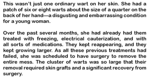
As described in this excerpt from James G. McCully’s Good Times In The Hospital, warts can be so extensive that they require a skin graft.

In the 19th century, skin grafts took many forms. By 1912 the process was down to a science, as detailed in Leonard Freeman’s Skin Grafting for Surgeons and General Practitioners. The basic method was established, with individual variations (“Donnelly, for some unexplained reason, prefers grafts from a portion of the skin subject to slight motion, such as the insertion of the deltoid muscle”), and complications and shortcomings were well known. (“Of all constitutional disorders, syphilis is probably the most disturbing, so much so that it has been claimed that grafting should never be attempted when this disease is present, and Freeman cites several cases which he thinks show that grafts will not adhere until syphilis has disappeared from the system.”) Many advancements had recently been made, even surprising doctors who didn’t quite expect them to work, and didn’t have a real theory to explain their success. The words of the late Dr. David Page (quoted here in 1872) were recalled with pity.

david-page-fatuity

Freeman’s book, like other contemporaneous reviews of skin grafting techniques, includes an extensive section on “anomalies in skin grafting”. This included inducing blisters and transplanting the thin blister roof; transplanting hairs (taking care to include the hair sheath); skin from a wide range of different animals, particularly frogs; bits of muscle; egg membrane; and of course, thin sections of rabbit testes.

E. Aievoli made use of thin sections of the testes of rabbits for purposes of grafting in four cases, assuming that the testicle possesses a greater cellular activity than other portions of the body. The results were undoubtedly good, but it does not follow that they were better than could otherwise have been obtained.

It’s obvious now that most of these substances would not fuse with, or become, actual skin, but they provided a suitable covering for the wound / ulcer / burn until it could heal. From a 1909 review in the Boston Medical & Surgical Journal, by Dr. Albert Ehrenfried:

Orcel (1888) states his belief that animal grafts act merely as a sort of protective dressing, and he is substantiated by the experiments of Beresowsky (1892).

Ehrenfried has a particular interest in converting surgeons to the “Reverdin method”, which sounds simple, safe, and advantageous in that the source of the graft is easy to obtain. You just take “morsels” of existing skin, aiming for a maximum of 1/8 inch in diameter (but cut from as deep as possible), and “seed” them over the “granulations of ulcers”. Held in place with “diachylon plaster”, they should allow healing to proceed faster, as it will spread from these new pieces of skin as well as from the margins of the ulcer.

Reverdin preferred this to the even easier “epithelial dust” method of Mangoldt.

method-of-mangoldt

And another procedure, that might make more sense than the early 20th century writers think, is sponge grafting, “introduced by Hamilton in 1881.”

Sponge does not grow fast to the surface as does skin, but acts merely as a stimulating support for the granulations, finally undergoing complete absorption. The procedure is much inferior in its results to the transplantation of cuticle, and is now seldom employed.

A fine Turkey sponge is selected, soaked in dilute nitrohydrochloric acid until all calcareous particles have been dissolved, and then placed for a time in desired in a solution of potassium hydroxid. Very thin slices, which are the most serviceable, can be cut … and sterilized by boiling, or in a 5 per cent solution of carbolic acid.

The sponge is then spread upon the granulations, which have been rendered as nearly aseptic as possible (see the method of Reverdin), and dressed much as if it were a transplantation of skin. The granulations soon acquire new energy and push their way into the interstices of the sponge, which often almost disappears beneath them, so luxuriant is the growth.

Providing an aseptic framework for tissue to grow around is something bioengineers do now, right? Albeit not with skin tissue.

* * *

But there’s one skin-grafting method that might have extra negative effects, beyond introducing infection, irritation, or just wasting everyone’s time. At the top of this piece, we see how skin grafts may be necessary for a bad case of warts. But what if you have a bad case of burns, from molten iron spilling into your boot? You also need a skin graft… and as a source of the new skin, you could use warts!

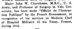
The first (and only?) example of this procedure was described by Dr. Charles Leale of New York in an 1878 report in The Medical Record. Presumably this was the same Charles Leale who 12 years earlier had attended at the Lincoln assassination, as a 23-year-old army doctor who would soon leave the public eye for many decades of lucrative obscurity. (Obscure enough that in some of the journals that reprinted his observations on wart grafting, he was credited as Charles Seale or Charles Lale.)

The Medical Record seems not to be archived in Google Books. The most comprehensive summary of Leale’s “The Use of Common Warts of the Hand in Skin-Grafting” now available is probably in the September 1878 Atlanta Medical and Surgical Journal, vol. XVI(6):354-355. Here’s an abstract, and then the whole thing. Why not, it’s only two pages.

As common warts of the skin are collections of vascular papillae, admitting of easy separation without injury to their excessively thick layer of well-nourished epidermis, the idea was conceived that, by their use for the purpose of skin-grafting, better and more rapid results would be obtained than when the ordinary skin of less vitality is used. As proof of  the theory, the following case is cited, where there had complete destruction of all the skin on the dorsum of the foot, involving to a great extent the deep cellular tissue, and where for several weeks no healing advanced until grafts of freshly removed warts from the patient’s hand immediately started little islands of tissue, which rapidly increased until they coalesced and met the margins of the border skin, thereby completely covering the foot by firm, protecting integument.

leale-warts-entire

Do we know if it’s possible to transplant warts and have them take root in new places? If it didn’t happen in this case, as Leale says it didn’t, it may be very hard to do.



Data update: How dyestuffs make stuff die

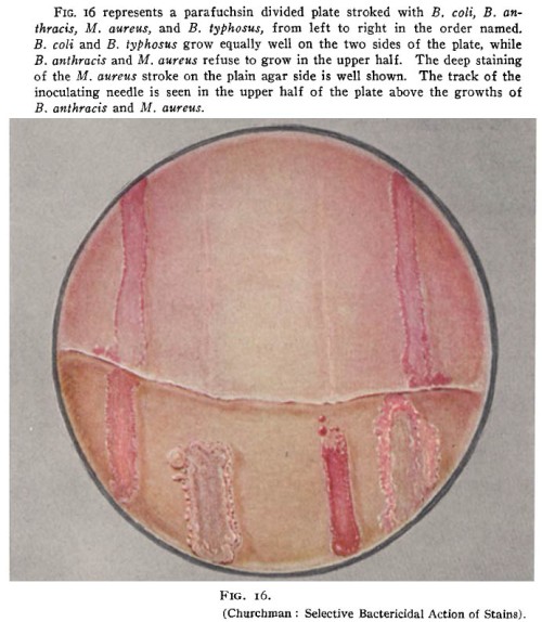
churchman-parafuchsin

This year, antibiotic researcher Mark Wainwright published a “Discussion” in the International Journal of Antimicrobial Agents, suggesting that we should take another look at a subject that he works on but most scientists lost interest in 6 decades ago: highly toxic synthetic molecules that can be used as local antiseptics and have distinctive bright coloration. What he calls “photoantimicrobials” represent a subset (those that can be targeted more precisely, because they are activated by light) of therapeutic molecules that earlier generations simply called flavines, anilins, or “dyes”.

Wainwright tells us:

Browning introduced the application of the basic dyes named above [acriflavine, crystal violet and brilliant green] to battlefield wounds in 1914/15. The aim was always to achieve antisepsis and healthy wound closure, and the approach was also used in pre-operative sterilisation. The dyes used thus represent local antiseptics. Importantly, however, they were an improvement on the available hypochlorite solutions owing to the fact that they were not inactivated by fluids associated with wounds, such as serum. Fleming’s rebuttal of this work, noted above, was based purely on laboratory measurement. However, the disinfection of real wounds and the concomitant recovery of patients could hardly be argued against.

By far the majority of clinical work involving dyes was carried out before 1945. At this juncture, the penicillins and other antibiotics or natural product-derived antibacterial agents became the driving force in infectious diseases therapy. From the distance of the early 21st century, it is apparent that these wonder drugs have not been used correctly and that their overuse has allowed a much more rapid development of resistance mechanisms than might otherwise have been the case.

In terms of modern healthcare, dye therapy might be considered obsolete and ineffective, and this would indeed be the case if the performance criteria were still the same as in Browning’s day. However, recent research has developed a different approach, one that uses locally applied dyes in conjunction with light to provide a microbial killing effect. Such dyes may be referred to as photodyes or, more properly, photosensitisers. Given their remarkable utility in the field under discussion, they are commonly called photoantimicrobials.

The photoantimicrobial effect has been known in the laboratory since the turn of the last century, but has only been investigated realistically since the early 1990s and is currently proposed foruse in oral and ENT (ear, nose and throat) disinfection. Its remarkable activity lies in the in situ production of reactive oxygen species (ROS) on illumination. The highly reactive nature of species such as singlet oxygen, the hydroxyl radical and the superoxide anion ensures rapid oxidative damage to simple cells, sufficient to guarantee cell death. More importantly, there are no known microbial resistance mechanisms able to combat ROS produced in this way.

Furthermore, among the lead compounds used in photo antimicrobial discovery are the same dyes used by Ehrlich and Browning; methylene blue, crystal violet and acriflavine.

Brilliantly colored solutions for treating localized infections and washing out wounds may be making a comeback. And not just to treat pets, for which acriflavine, for example, is now used. In preparation for that retro craze, let’s look at some of the original pre-Depression research. churchman-gazette

Most of it is in German, which I will ignore.

Much of the non-German work on “dye therapy” was led by urologic surgeon John W. Churchman of Yale University. At the top of this post is a rare color photo of bacteria in a Petri dish from the time when Julius Richard Petri (1852-1921) was still alive. This comes from a 1913 Journal of Experimental Medicine paper by Churchman, The Selective Bactericidal Actions of Stains Closely Allied to Gentian Violet, whose figures are otherwise black & white. Even the limitless moneybags of Old Elihu and the Rockefeller Institute only extended to printing one color figure.

* * *

Churchman’s papers are not very quantitative, but other “dye therapy” researchers did large-scale comparisons of multiple colored antibiotics, at multiple concentrations, against multiple bacterial species.

krumwiede-title

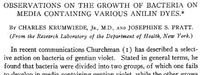
On January 1, 1914, the Journal of Experimental Medicine published the Observations of Josephine S. Pratt and Charles Krumwiede Jr. (not to be confused with their Further Observations, published 4 months later). They established a method for testing large numbers of drug/bacteria combinations while minimizing the amount of agar needed, by putting two samples in each Petri dish, slanted away from each other. Here they describe the process in vague terms that would be better presented as a 5-minute video.

As a routine a batch of agar was selected which was found especially suitable for the more feebly growing [bacteria]. To the hot agar an appropriate volume of a watery solution of dye was added to give the final dilution desired. The same agar was used in each experiment.

The most convenient and economical method was found to be as follows. One unopened Petri dish was used to tilt up one side of a second dish. In the opposite side was poured just sufficient agar to give a satisfactory slant. This dish was covered and used to tilt up a third dish, and so on in a row. After the agar had set, slants were poured in the other side of the Petri dishes which were tilted in the reverse direction. In this way two mixtures of agar could be used in the same dish, very little agar being required for each slant.

After inoculating all these plates with bacteria, they used the eye test. Compared to a control plate with no dyestuffs, how much did the dye prevent the bacteria from growing after 18 to 24 hours? Not at all? Was it “restrained”? “Markedly restrained”? Completely eliminated?

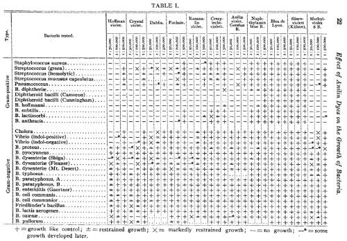
Here’s Table 1.

dyestuffs-original-table-1

That is a lot of information. 11 drugs, tested against 30 bacteria (12 Gram-positive and 18 Gram-negative), makes 330 combinations, of which only 7 potential tests were not performed. (I think the ellipsis means “not performed”.) Each of these 323 tests was done at 3 concentrations, making 969 data points in a single table.

To turn this into graphs would require many graphs. It would also require us to turn the X’s and +’s into a semiquantitative system (let’s say + = 4, ± = 3, X = 2, —* = 1, and — = 0). And since each data point is restricted to those 5 possibilities, you wouldn’t gain much by looking at a bunch of individual dots or bars anyway.

Let’s leave it as a table.

The main flaw of this table is that all the symbols look similar, except “—” . The bottom half of the table is a wall of symbols for different degrees of growth. It’s hard to see trends, because the symbols are hard to distinguish from each other, so it looks like every bacterium is resistant to every drug. Which is not quite true.

Instead of these text symbols, how about something that may be more intuitive — representing the 5 levels of growth as 5 colors. This may not be intuitive in every culture, and it may not work for the color-blind or in B&W printouts, but I took the liberty of changing the table. Now as bacteria proliferate, they pass from “no growth” (white) through yellow, green, and purple stages until they attain “growth like control” (charcoal grey). At first I wanted to range from white to black, but if the cells are black you couldn’t see the lines between them. Or maybe you can’t see them anyway.

For the tests that weren’t done, I replaced the ellipses with neutral grey. And finally, the two types of “diphtheroid bacilli” produced exactly the same results, so they were conflated into one.

dyestuffs-table-1

So where does this data lead us? Krumwiede and Pratt draw limited conclusions. The tables speak for themselves. To briefly summarize their Summary section: the “streptococcus-pneumococcus group” is more dye-resistant, and the “dysentery bacillus group” is highly unpredictable with dye-resistance showing “no correlation with the common differential characteristics”. Also, they discovered mutations that make bacteria lose resistance. (“Among Gram-negative bacteria a strain is occasionally encountered which will not grow on violet agar, differentiating it from other members of the same species or variety.”) Finally, in the middle of the Summary section they say this.

The reaction is quantitative, although the quantitative character is more marked with some species than with others.

Now, a modern reviewer would look unkindly on that sort of admission. First, it seems like it’s not quantitative, it’s semiquantitative. Instead of measuring the number of colonies, you’re grading “growth” on a scale from 1 to 5. And which are the species that don’t have a “quantitative character”? Why don’t you use some other method to see how well those ones are growing? And by the way, how repeatable is your eye test? And what does it mean exactly? Let’s see examples of “growth like control”, “restrained growth”, and “markedly restrained growth”. Does it mean there were fewer colonies, or the colonies were smaller, or both? And why use the same 3 concentrations of all 11 drugs? Maybe 1:500,000,000 would have still killed the bacteria, and be less toxic to patients.

That’s what I’d say, if the journal hadn’t told me “Your review is now 100 years late and consequently is no longer needed.”

* * *

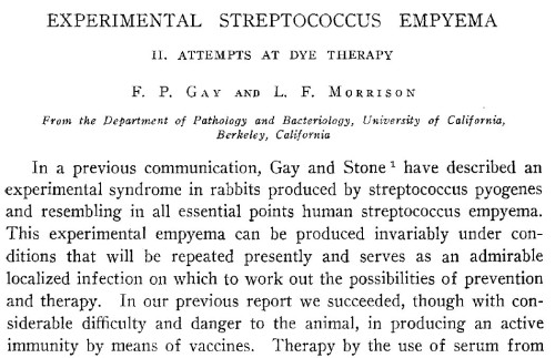
One more paper: from 7 years later, Gay and Morrison in the January 1921 Journal of Infectious Diseases. Whereas Krumwiede & Pratt (1914) was the first in their series, this is a sequel.

gay-morrison-title

Krumwiede & Pratt took a limited range of dyes, and used then on every sort of bacteria they could find. Now Gay & Morrison use every dyestuff they can find on a limited range of bacteria. This is a much longer paper, so I’ll just address their first set of data, about bacterial growth in culture, and ignore their innovative rabbit empyema model.

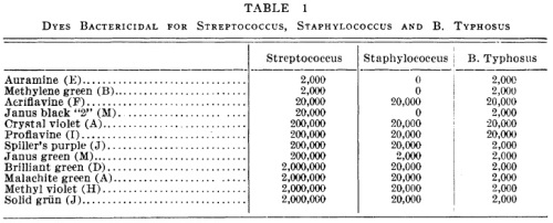
Table 1:

gay-morrison-table-1

In this table, the numbers mean the reciprocal of the minimum inhibitory concentration (MIC). The MIC is the smallest concentration of the drug that will prevent bacteria from growing. If the MIC is 5 ng/ml, the bacteria should grow if the concentration is lower than that, and the bacteria should die if the concentration is 5 mg/ml or greater.

“2,000” means a 1 to 20,000 dilution. I think that’s weight/volume (1 gram in 20,000 milliliters). The lower the number in this graph, the more concentrated the drug needs to be to kill bacteria. A drug marked “2,000” needs to be a thousand times more concentrated to have the same effect as a drug marked “2,000,000”.

This table is clear. Intuitively we look at the numbers and recognize that 2,000 is smaller than 20,000 and therefore represents less of a dilution. If they were in scientific notation, it wouldn’t be as intuitive.

The only problem is the use of “0”. The most concentrated dyes they used were at a 1:2,000 concentration. If the bacteria still grew, they listed this drug as “0”, or not active against the bacteria. Taken literally, “0” means Staphylococcus would grow on media consisting entirely of methylene green. I would just write the word “inactive” instead of the number 0.

* * *

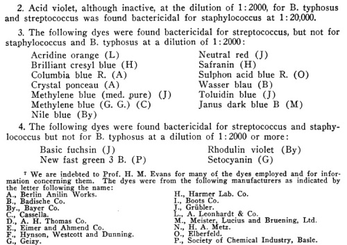
I might also expand the table to include the rest of the 40 dye molecules Gay & Morrison used in this study. That’s right — in addition to the 12 molecules listed above, they have data on Acid fuchsin, Acid violet, Acridine orange, Azo-acid red, Basic fuchsin, Benzo-azurin, Brilliant cresyl blue, Columbia blue R, Congo red, Crystal ponceau, Cyanin B, Diamil blue, Erioglaucin A, Janus dark blue B, Methylene blue (medical purity), Methylene blue GG, Neutral red, New fast green 3B, Nile blue, Oxamin violet, Rhodulin violet, Safranin, Sauer grün, Scarlet 6R, Setocyanin, Sulphon acid blue R, Toluidin blue, and Wasser blau.

But most of the data is in paragraph form. And it’s not immediately clear.

gay-morrison-excerpt

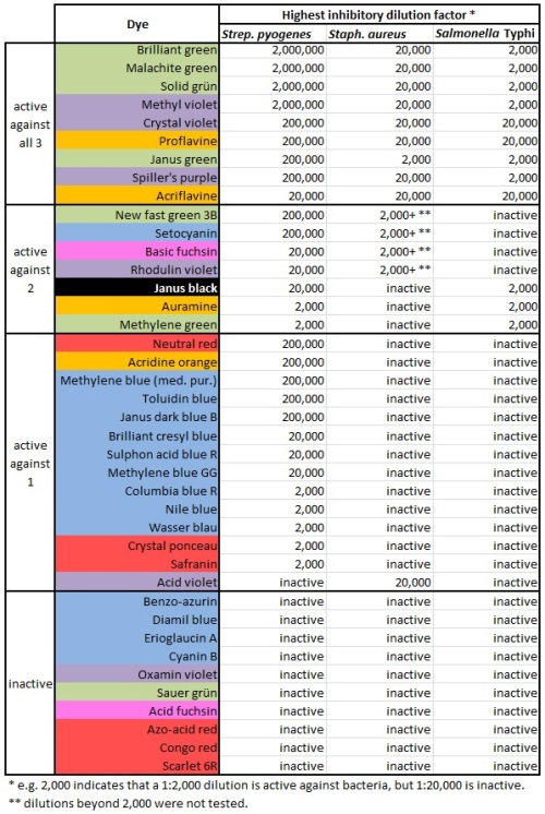
Let’s make an expanded version of the table above, containing all the dyestuffs. Even the dyestuffs that never killed any of the bacteria. And make some other changes:

  • Keep them arranged in order of effectiveness, but put the most effective ones at the top.
  • In fact, divide them into categories. Those that inhibited all 3 bacterial species, those that inhibited 2, those that inhibited 1, and those that were completely ineffective.
  • Update the nomenclature. We consider Bacillus typhosus to be Salmonella now.
  • And… why not color-code the chart. Maybe blue dyes will do one thing, and red dyes will do something else. Might as well include that information. Patterns may emerge.

gay-morrison-table-2

Isn’t it nice to see those colors? And it shows how much easier it is to kill Streptococcus pyogenes. There’s only one dye that kills either Staphylococcus or Salmonella but fails to kill S. pyogenes. And it shows that blue and red dyes aren’t very antibacterial, while green ones kill bugs dead. Why is that?

Medical privacy, 1950 style

Without really working in a medical field, I still hear a lot about medical privacy and HIPAA. And how any public presentation of patient information needs to be anonymized, any identifying details need to be removed, any irrelevant personal details need to be removed, populations need to be profiled rather than individuals, and generally all publications about human patients must run through a battery of administrators to make sure everything is in compliance with federal rules, state rules, institutional rules, the institutional rules of other institutions that collaborated with us, and heaven knows what else.

Not so long ago, the standards were different. Today we’ll look at a 1950 article on “Primary Cutaneous Cryptococcosis” from Archives of Dermatology and Syphilology (later renamed Archives of Dermatology, and then JAMA Dermatology). An actual doctor could explain what parts of this article would be scrubbed now, and what parts are still OK. I don’t really know. But what struck me was that nearly all the personal details seem unrelated to the actual topic of the article. Meanwhile, author William M. Gandy, M.D. points out that “family history was irrelevant”. Indeed, it would be even more irrelevant to point out the patient’s mother’s alcoholism, or grandfather’s yaws and phossy jaw.

gandy-title

Information we can glean from the piece:

– “The patient had been under continuous observation and treatment at the Charity Hospital of New Orleans since July 2, 1945. Successive complaints brought her at one time or another to the attention of the medical, surgical, gynecologic and dermatologic services.”

– “She stated that she had been ‘anemic’ and afflicted with ‘enlarged glands’ since childhood. She had measles, mumps, chickenpox and scarlet fever in early life.”

– She was “first seen by us” (the dermatology clinic) on August 14, 1947, at age 29.

– She was white and a lifetime resident of New Orleans, Louisiana.

– At this time she was divorced with one child.

– That child was conceived in 1942 in Cleveland, Ohio, but born in New Orleans.

– For months after the birth of the child, she was given weekly X-ray treatments for “extensive” genital warts. The “Roentgen rays” did no good and she was then given podophyllin ointment, which led to “a severe burn followed by a slough of much of the vulva”.

– During 1942 she was diagnosed with Boeck‘s sarcoid.

– During 1947 she had stones removed from her right kidney.

– During 1947 she underwent a vulvectomy and a ureterotomy.

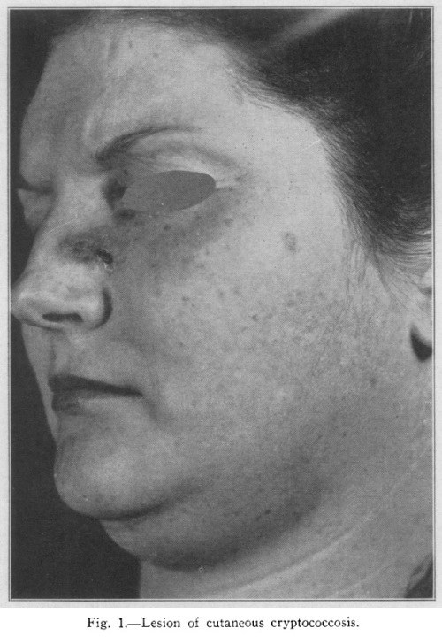
– The whole point of this paper, a cutaneous Cryptococcus infection on her face, was probably the least of her problems and had little to do with all this other stuff anyway.

– Very unusual for cryptococcosis, this infection was limited to a patch of skin and not found in the blood, cerebrospinal fluid, urine, sputum or mucosal swabs.

– 12 mice were injected with half a milliliter of a broth culture of her strain of Cryptococcus. 6 of the mice died within 12 days, and the remaining 6 died within 21 days.

– Between the end of 1947 and April 1949 she left Charity Hospital, married her second husband, and returned to Charity Hospital, this time the urology clinic, after developing a renal abscess and uremia. This was not a Cryptococcus infection.

– “Further physical and laboratory investigations were not possible at the time because of the patient’s uncooperative attitude and the severity of her illness.”

– Finally… here’s a picture of her face.

gandy-cryptococcus-1950

If you know this woman, you may be glad to know that she received a large amount of medical care free from top clinicians, as Charity Hospital was adjacent to the LSU Health Sciences Center. Was this care better than what would have been provided in 1736, when the hospital was founded? Of course! What kind of question is that? Or anyway, I think so, but I’m no historian.

Gallery: The amazing ink-proof yeast capsule

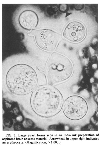
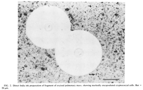
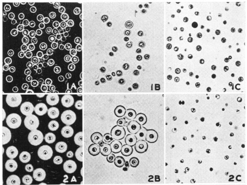
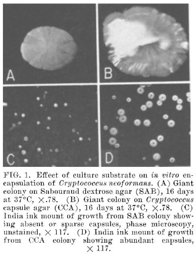
Observed doctors and medical students as they learn about the workings of the clinical microbiology lab, I’m impressed by their love of the India ink test for cryptococcus. The way this test works is: Cryptococcus is a type of infectious yeast that looks a lot like Candida if you just do a gram stain. But it has a polysaccharide capsule around each cell (unless for some odd reason it isn’t producing a capsule), wider than the cell itself. So if you put Cryptococcus in a colored liquid, most famously a solution of India ink, the polysaccharide capsule shows up as a huge empty white area around the cell. Whereas with Candida, only the cell itself is white.

We apparently don’t use this test regularly anymore, but we still show it to people in case they need to know what it is.

Something about the India ink test just makes people happy. A lot of diagnostic microbiology uses techniques that were developed several generations ago, but this one is just so simple, requiring not “acid alcohol” or various toxic red and purple substances, but merely the simplest form of ink, developed millennia ago. And to use the phrase “India ink”, instead of “colloidal carbon” or something, is such an anachronism in the 21st century. Most of us last saw that phrase when reading some classic of literature like The Secret Garden or A Bear Called Paddington. And aside from the name, there’s something magical about seeing this invisible capsule appear around what seemed to be a normal yeast cell. Like lemon-juice ink made visible.

* * *

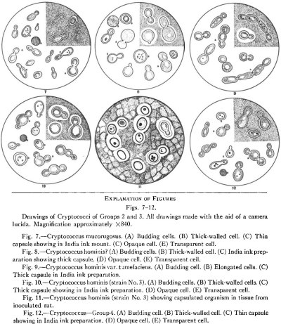
So here are some depictions of India-ink-stained Cryptococcus in the literature. First, camera lucida drawings from a 1935 JID paper by Rhoda W. Benham (Cryptococci — their identification by morphology and serology) that must have been a handy field guide to Cryptococcus species. The top right corners of the dishes are shaded to show how they look under India ink.

crypto-benham

* * *

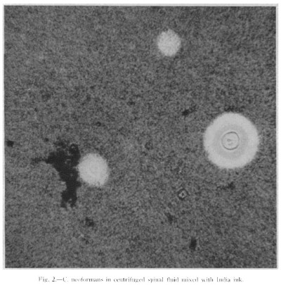
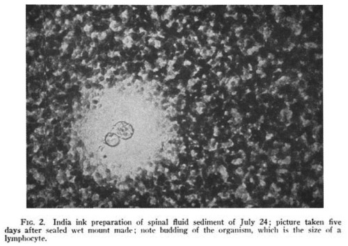
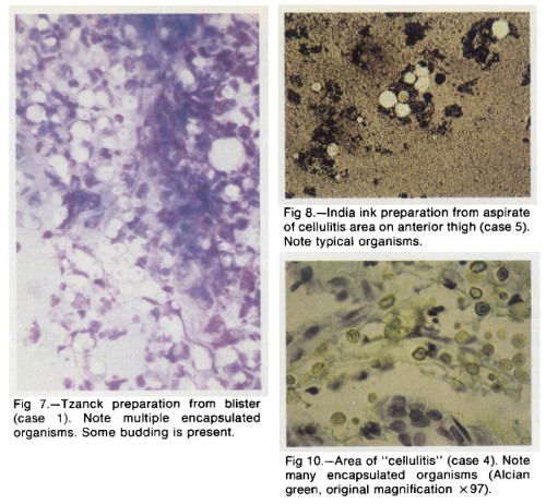
Now, some photos of patient tissues directly stained with India ink.

From Wilson HM, Duryea AW (1951), Cryptococcus meningitus (Torulosis) treated with a new antibiotic, actidione®. Archives of Neurology & Psychiatry 66(4):470-480.

crypto-duryea

* * *

From Carnecchia BM, Kurtzke JM (1960), Fatal toxic reaction to amphotericin B in cryptococcal meningo-encephalitis. Annals of Internal Medicine 53(5):1027-1036.

crypto-carnecchia

* * *

From Schupbach CJ, Wheeler CE Jr, Briggaman RA, Warner NA, Kanof EP (1976), Cutaneous manifestations of disseminated Cryptococcosis. Archives of Dermatology 112(12):1734-1740. Note “Tzanck preparation”, looking for multinucleated giant cells.

crypto-schupbach

* * *

From Love GL, Boyd GD, Greer DL (1985), Large Cryptococcus neoformans isolated from brain abscess. Journal of Clinical Microbiology 22(6):1068-1070.

crypto-love-1985

* * *

From Bottone EJ, Kirschner PA, Salkin IF (1986), Isolation of highly encapsulated Cryptococcus neoformans serotype B from a patient in New York City. Journal of Clinical Microbiology 23(1):186-188.

crypto-bottone

* * *

And some images of cells grown in culture. Ending with one in color!

From Neill JM, Abrahams I, Kapros CE (1950), A comparison of the immunogenicity of weakly encapsulated and of strongly encapsulated strains of Cryptococcus neoformans (Torula histolytica). Journal of Bacteriology 59(2):263-275.

crypto-neill

* * *

From Littman ML, Tsubura E (1959), Effect of degree of encapsulation upon virulence of Cryptococcus neoformans. Proceedings of the Society for Experimental Biology & Medicine 101:773-777.

crypto-littman

* * *

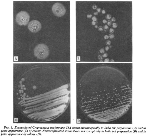
From Bulmer GS, Sans MD, Gunn DM (1967), Cryptococcus neoformans I: Nonencapsulated mutants. Journal of Bacteriology 94(5):1475-1479.

crypto-bulmer

* * *

From Dykstra MA, Friedman L, Murphy JW (1977), Capsule size of Cryptococcus neoformans: Control and relationship to virulence. Infection & Immunity 16(1):129-135.

crypto-dykstra

* * *

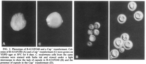
From Chang YC, Kwon-Chung KJ (1994), Complementation of a capsule-deficient mutation of Cryptococcus neoformans restores its virulence. Molecular & Cellular Biology 14(7):4912-4919.

crypto-chang

* * *

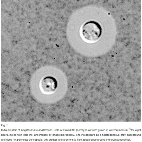
From Doering TL (2000), How does Cryptococcus get its coat? Trends in Microbiology 8(12):547-553.

crypto-doering

* * *

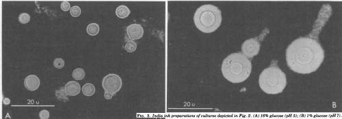
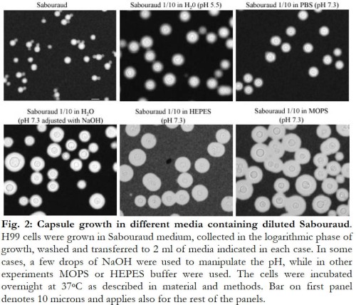
From Zaragoza O, Casadevall A (2004), Experimental modulation of capsule size in Cryptococcus neoformans. Biological Procedures Online 6(10):10-15.

crypto-zaragoza-2d

* * *

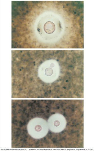
From Zerpa R, Huicho L, Guillén A (1996), Modified India ink preparation for Cryptococcus neoformans in cerebrospinal fluid specimens. Journal of Clinical Microbiology 34(9):2290-2291.

crypto-zerpa

* * *

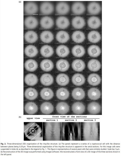
And a bonus: High-tech 3-dimensional visualization! These are 40 focal “slices” of a single cell. From Zaragoza O, McClelland EE, Telzak A, Casadevall A (2006), Equatorial ring-like channels in the Cryptococcus neoformans polysaccharide capsule.

crypto-zaragoza-3d